
Chapter 17 – Normal, Reference Images, Unlabelled and Labelled
Chest
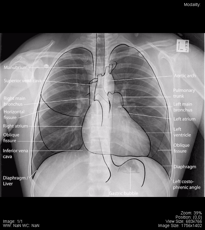
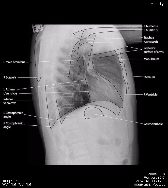
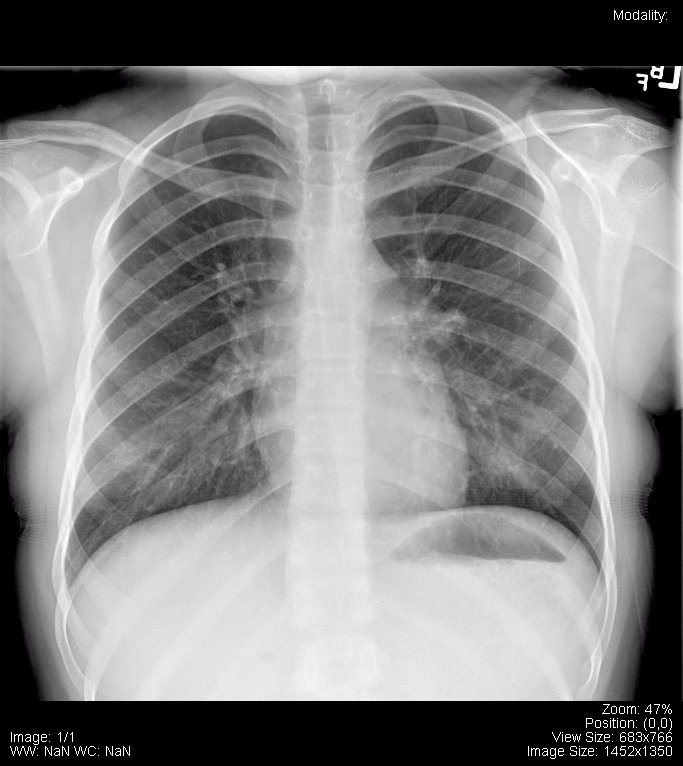
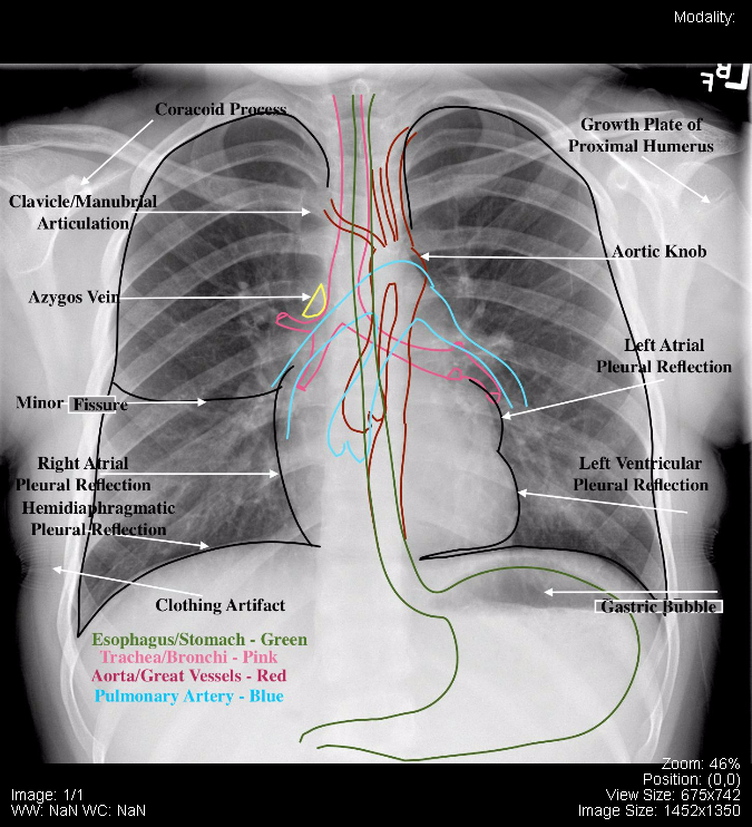
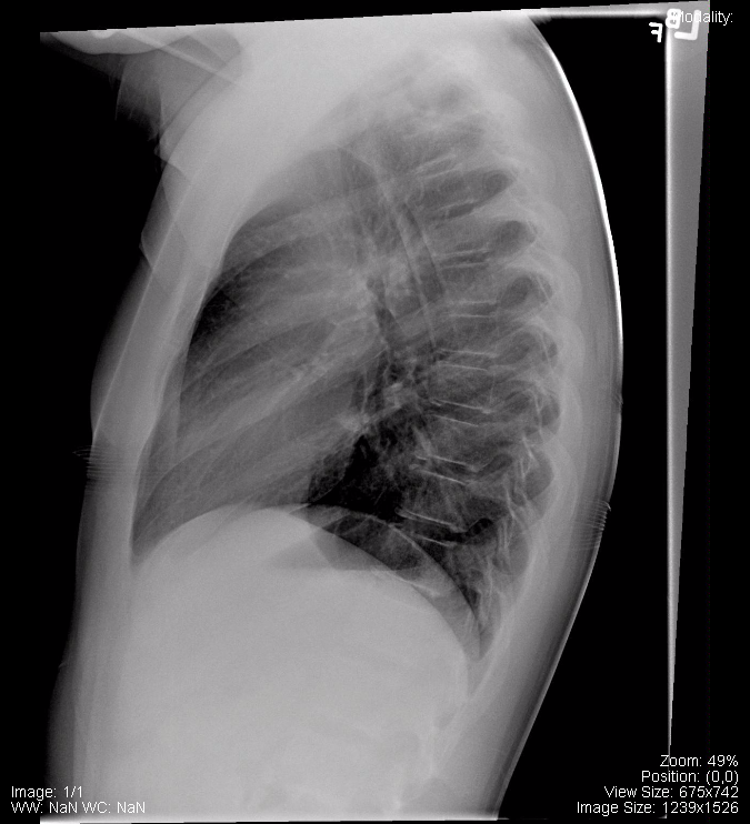
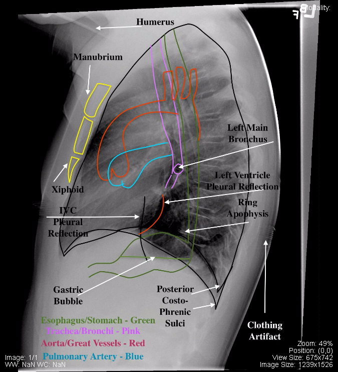
The following are normal PA and Lateral x-ray images of the chest:
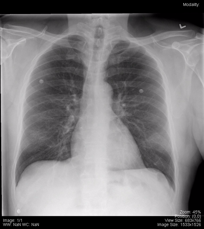
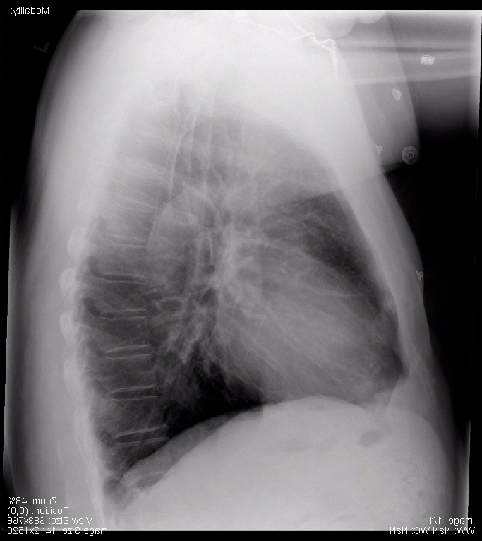
ODIN Link to Unlabelled Chest x-ray Images – https://mistr.usask.ca/odin/?caseID=20170402103011944
ODIN Link to Labelled Chest x-ray Images – https://mistr.usask.ca/odin/?caseID=20170103165555291
The following is a normal AP (Portable) x-ray of the chest:

ODIN Link to Normal AP (Portable) Chest x-ray – https://mistr.usask.ca/odin/?caseID=20170114100724626
The following are normal pediatric chest x-rays:
ODIN Link to Images – https://mistr.usask.ca/odin/?caseID=20170825125714420
The following are sections from a High Resolution CT (HRCT) of a normal chest:
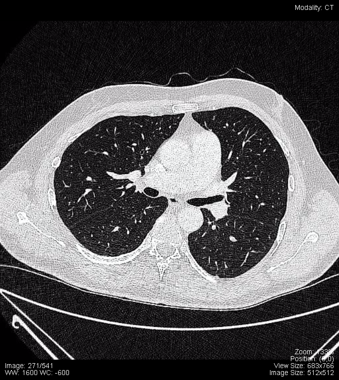
ODIN Link to HRCT Images – https://mistr.usask.ca/odin/?caseID=20170731230443139
The following is a normal CT Angiography (CTA) of the Pulmonary Arteries:


ODIN Link to CTA – https://mistr.usask.ca/odin/?caseID=20170803170331806
Attributions
All figures in “Chapter 17: Chest” by Dr. Brent Burbridge MD, FRCPC, University Medical Imaging Consultants, College of Medicine, University of Saskatchewan is used under a CC-BY-NC-SA 4.0 license.
