Want to create or adapt books like this? Learn more about how Pressbooks supports open publishing practices.
15. Reproduction
(Figures only, for the moment!)
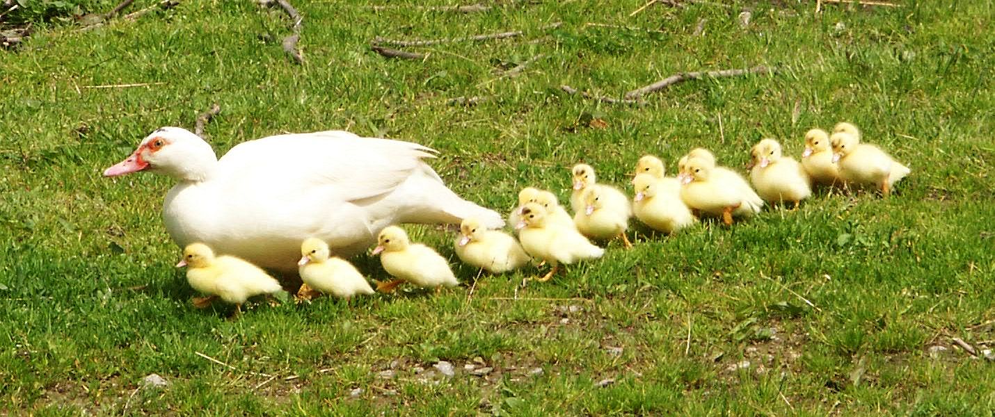
Figure 15.x Reproduction is the process of making copies of yourself. Do you have all your ducks in a row?
Photo: Georg Mittenecker, https://commons.wikimedia.org/wiki/File:Duckfamily.jpg CC BY-SA 2.5, 2006.
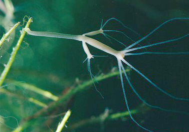
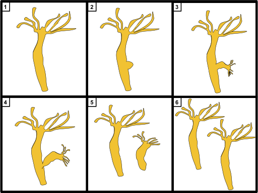
Figure 15.x Some animals reproduce by budding. Cell division in one part of the body produces a group of undifferentiated cells that forms a bud. The bud develops into a miniature individual that grows into an adult. Individuals produced by budding are clones – in other words, genetically identical to their parent.
Photo: Lifetrance / en.wikipedia, https://commons.wikimedia.org/wiki/File:Hydra_oligactis.jpg CC BY-SA-3.0, 2009; Diagram: A.houghton19 / Wikimedia Commons, https://commons.wikimedia.org/wiki/File:Hydra_Budding.svg CC BY-SA-4.0, 2018.
Figure 15.x A catshark egg with a developing foetus inside. You can see where the foetus is getting its supply of nutrients: the yolk and the umbilical cord connecting the yolk to the body. Animals that lay eggs are said to be oviparous.
Photo: Sander van der Wel, https://commons.wikimedia.org/wiki/File:Scyliorhinus_canicula_foetus_in_an_egg.jpg CC BY-SA 2.0, 2010.
Figure 15.x An aphid giving birth. Animals that give birth instead of laying eggs are said to be viviparous. Many aphids are live-bearing (viviparous) and egg-laying (oviparous) during different parts of the their life cycle. Amazingly, the daughter aphid being born in this photo may already be pregnant: Many aphid species reproduce apomictic parthenogenesis during the viviparous part of their life cycle. In apomictic parthenogenesis, the germ cells skip meiosis during development into egg cells (oocytes), such that the daughters are clones of their mother.
Photo: MedievalRich / Wikimedia Commons, https://en.wikipedia.org/wiki/File:Aphid-giving-birth.jpg CC BY-SA 3.0, 2007.
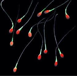
Figure 15.x Human sperm cells. In this fluorescence image, DNA is stained red, and the rest of the cell is stained green.
Fluorescence micrograph: Gilberto Santa Rosa, https://commons.wikimedia.org/wiki/File:Sperm-20051108.jpg CC BY 2.0, 2005.
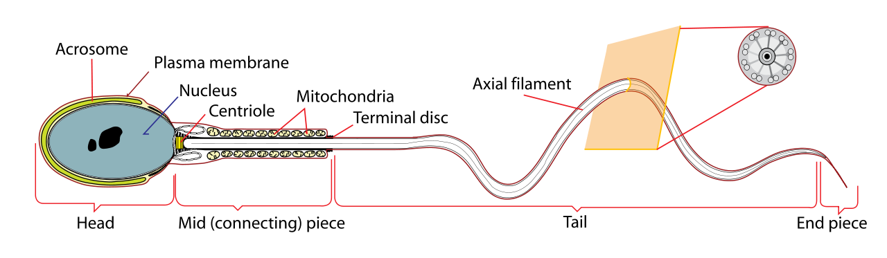
Figure 15.x Diagram of a sperm cell. Do you know what each part does?
Diagram: Mariana Ruiz, https://en.wikipedia.org/wiki/File:Simplified_spermatozoon_diagram.svg public domain, 2009.
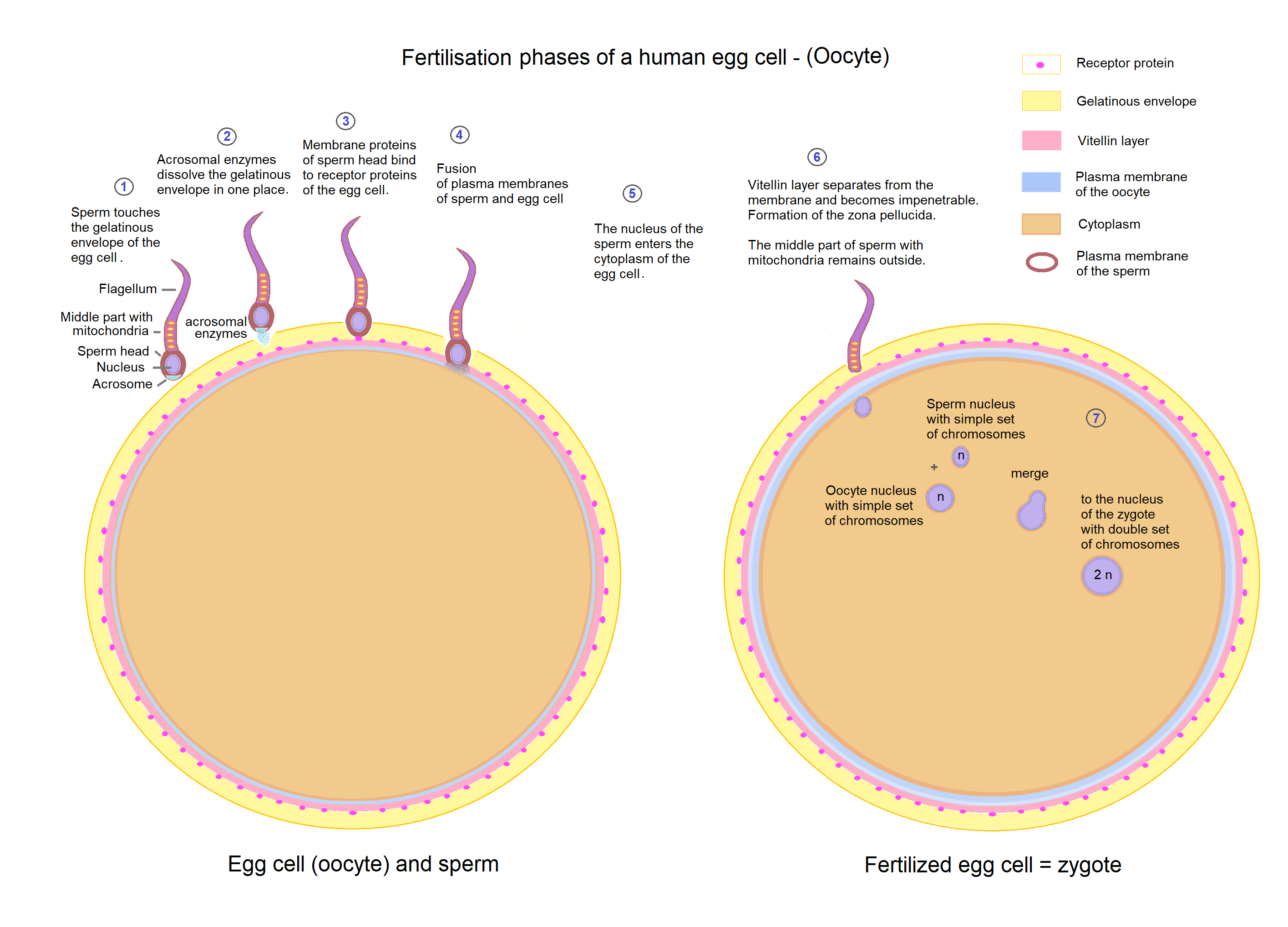
Figure 15.x Fertilisation.
Diagram: Sciencia58 / Wikimedia Commons, https://commons.wikimedia.org/wiki/File:Egg_cell_fertilization_-_Zygote.png public domain, 2019.
Figure 15.x Cross-section of seminiferous tubules of a rabbit testicle. Can you see the developing sperm cells?
Photo: James Scott, https://commons.wikimedia.org/wiki/File:Rabbit_testis.jpg public domain, 2016.
Figure 15.x (a) Sperm production through meiosis. (b) Anatomy of seminiferous tubules in a mammalian testis.
Diagram: Anatomy and Physiology, Rice University; Access for free at https://openstax.org/books/anatomy-and-physiology/ CC BY 4.0, 2018.
Figure 15.x Production of steroid hormones from cholesterol in vertebrate animals.
Diagram: David Richfield and Mikael Häggström, “Diagram of the pathways of human steroidogenesis”WikiJournal of Medicine 1 (1), DOI:10.15347/wjm/2014.005, ISSN 20024436 CC BY-SA 3.0, 2014.
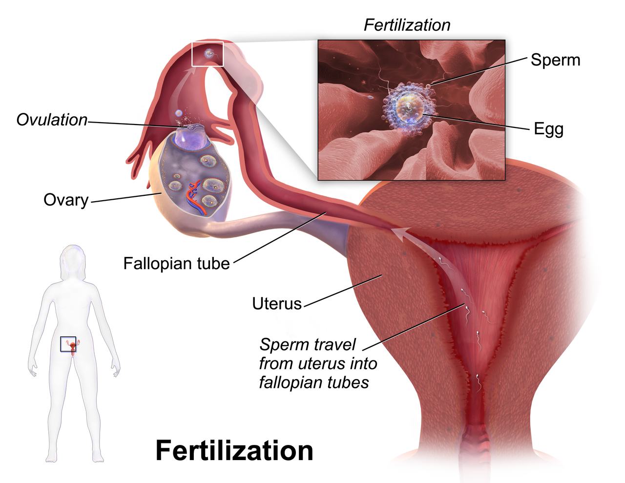
Figure 15.x Fertilisation in the human body.
Source: Blausen.com staff, Medical gallery of Blausen Medical 2014, WikiJournal of Medicine 1 (2), DOI:10.15347/wjm/2014.010, ISSN 2002-4436 CC BY 3.0, 2014.
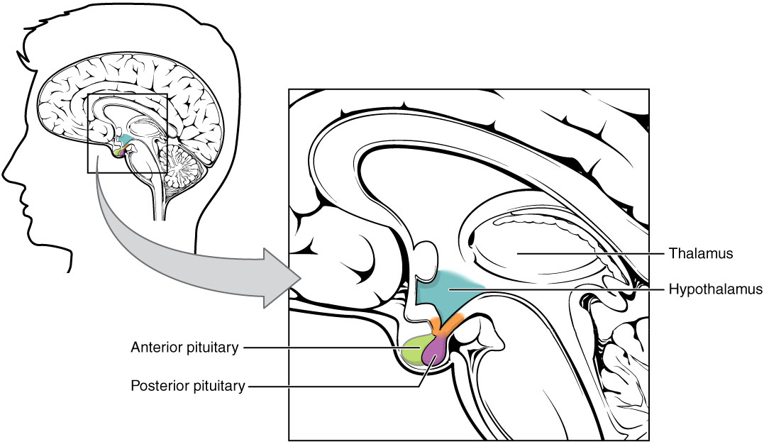
Figure 15.x In vertebrate animals, the hypothalamus, posterior pituitary, and anterior pituitary are structures near the base of the brain that produce and secrete several hormones that are important for reproduction, among many other functions.
Diagram: Adapted from Anatomy and Physiology, Rice University; Access for free at https://openstax.org/books/anatomy-and-physiology/ CC BY 4.0, 2018.
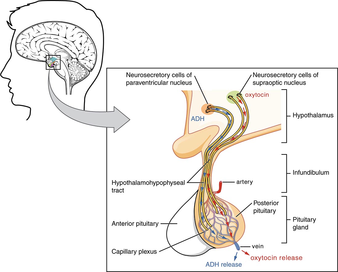
Figure 15.x The posterior pituitary is derived from nervous tissue, and contains the axon ends (terminals) of hormone-producing neurons in the hypothalamus. These special neurons are called “neurosecretory cells” because their axons don’t end in a synapse, but instead release their signalling molecules as hormones into a web of blood capillaries. Shown here is production of the peptides antidiuretic hormone (ADH) and oxytocin in neuron cell bodies in the hypothalamus, and release of these two hormones into the blood in the posterior pituitary.
Diagram: Adapted from Anatomy and Physiology, Rice University; Access for free at https://openstax.org/books/anatomy-and-physiology/ CC BY 4.0, 2018.
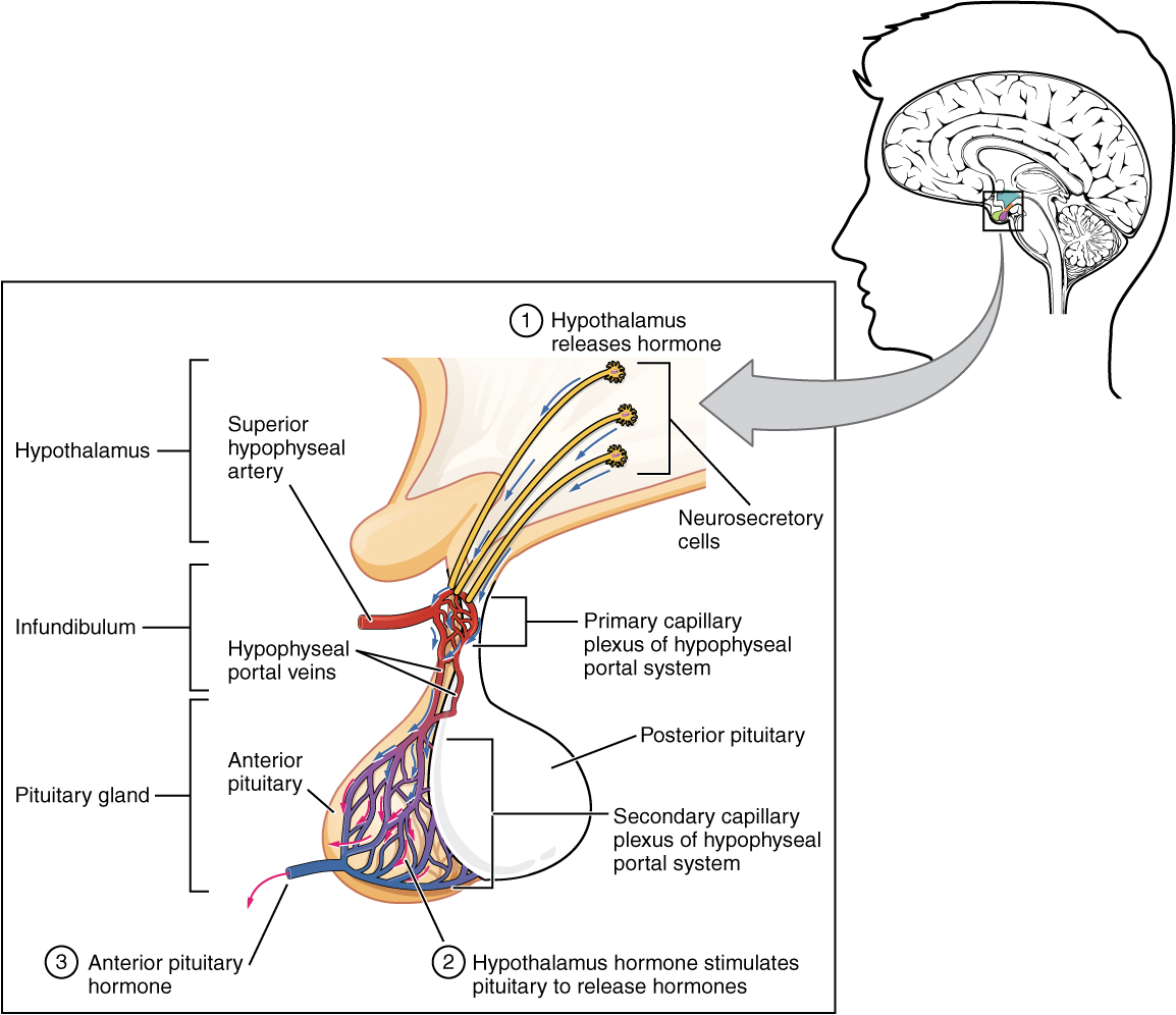
Figure 15.x The anterior pituitary is derived from epithelial tissue, and produces and releases hormones in response to releasing hormones from the hypothalamus. Blood in a portal vessel system carry the releasing hormones from the edge of the hypothalamus to the anterior pituitary, which produces hormones for general circulation.
Diagram: Anatomy and Physiology, Rice University; Access for free at https://openstax.org/books/anatomy-and-physiology/ CC BY 4.0, 2018.
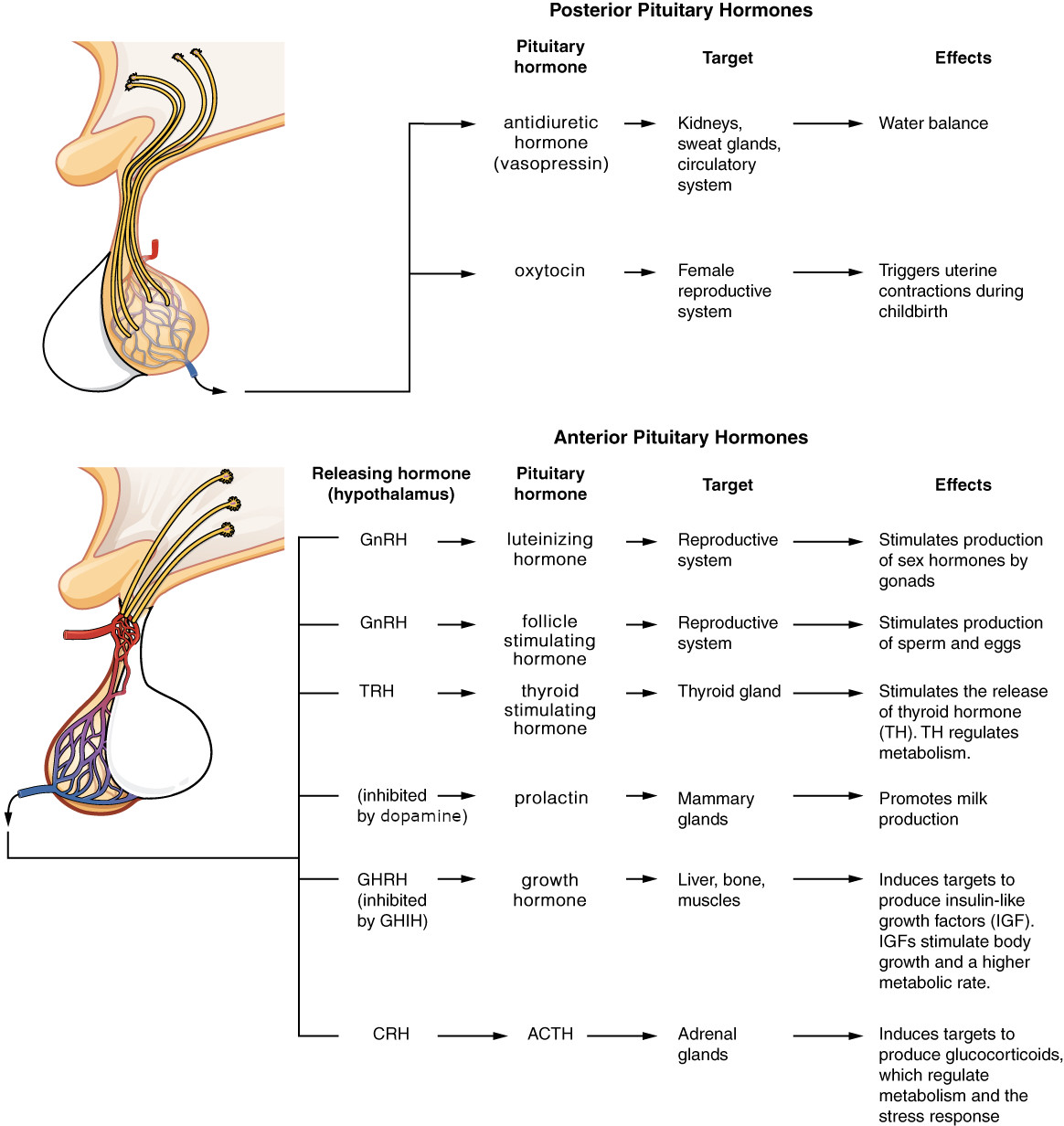
Figure 15.x Summary of anterior and posterior pituitary function.
Diagram: Adapted from Anatomy and Physiology, Rice University; Access for free at https://openstax.org/books/anatomy-and-physiology/ CC BY 4.0, 2018.
Diagram and caption from: Anatomy and Physiology, Rice University; Access for free at https://openstax.org/books/anatomy-and-physiology/ CC BY 4.0, 2018.
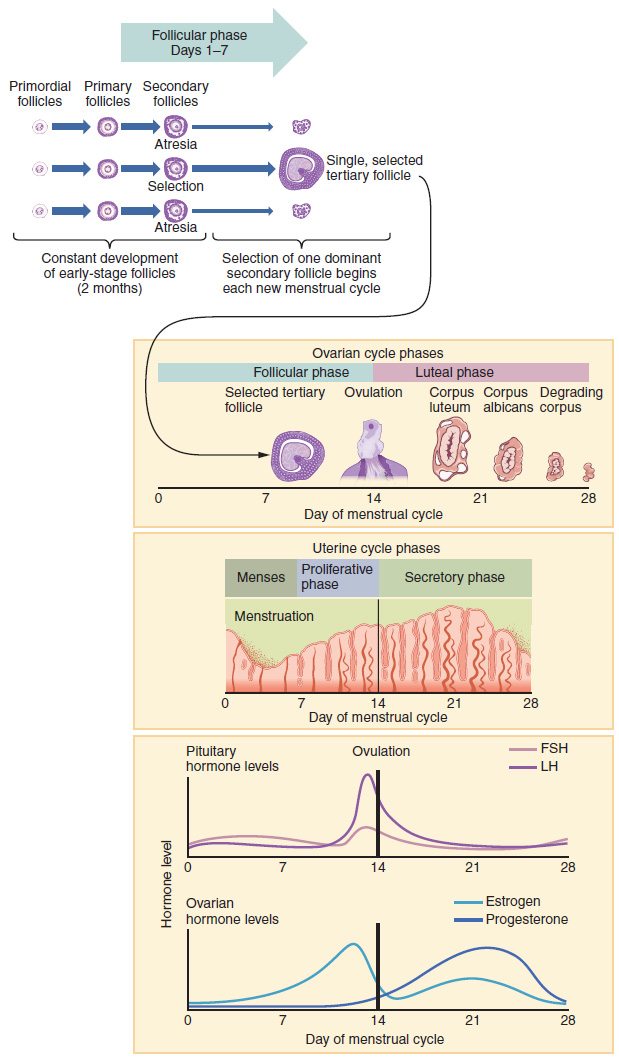
Figure 15.x The ovarian cycle in humans.
Diagram: Adapted from Anatomy and Physiology, Rice University; Access for free at https://openstax.org/books/anatomy-and-physiology/ CC BY 4.0, 2018.
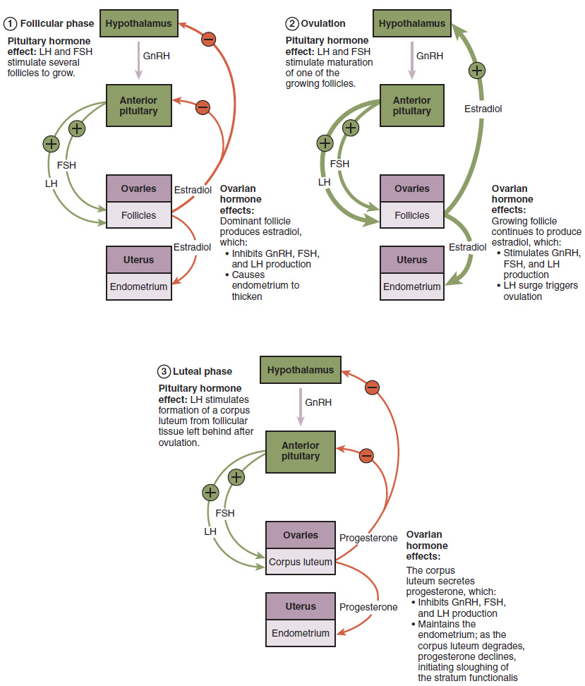
Figure 15.x Hormonal signalling during the three main stages of the ovarian cycle in humans. Can you identify the positive and negative feedback loops?
Diagram: Anatomy and Physiology, Rice University; Access for free at https://openstax.org/books/anatomy-and-physiology/ CC BY 4.0, 2018.
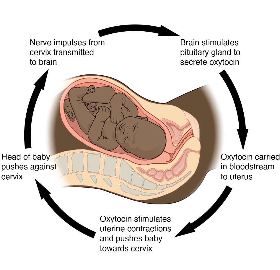
Figure 15.x Childbirth (parturition) is a classic example of a positive feedback loop. When the baby’s head pushes against the cervix (1), it stimulates nerve impulses from the cervix to the brain (2). The grain responds by signalling to the pituitary gland to release a hormone called oxytocin into the bloodstream (3). The blood then carries the oxytocin to the uterus (4) causing uterine muscle contractions (5), the push the baby harder against the cervix (1), stimuating more frequent nerve impulses to brain (2), and so on.
Diagram on left/top: Biology, Rice University; Access for free at https://openstax.org/books/biology-2e/pages/1-introduction CC BY 4.0, 2018.
Figure 15.x The “let-down” reflex in mammalian lactation.
Diagram: Biology, Rice University; Access for free at https://openstax.org/books/biology-2e/pages/1-introduction CC BY 4.0, 2018.