Case Studies
Case 1: Dolly
Dolly, a 9-year-old Quarter horse mare, has a 3 day history of nasal discharge and decreased appetite after being trailered from Texas. Now inappetent, febrile, and depressed.
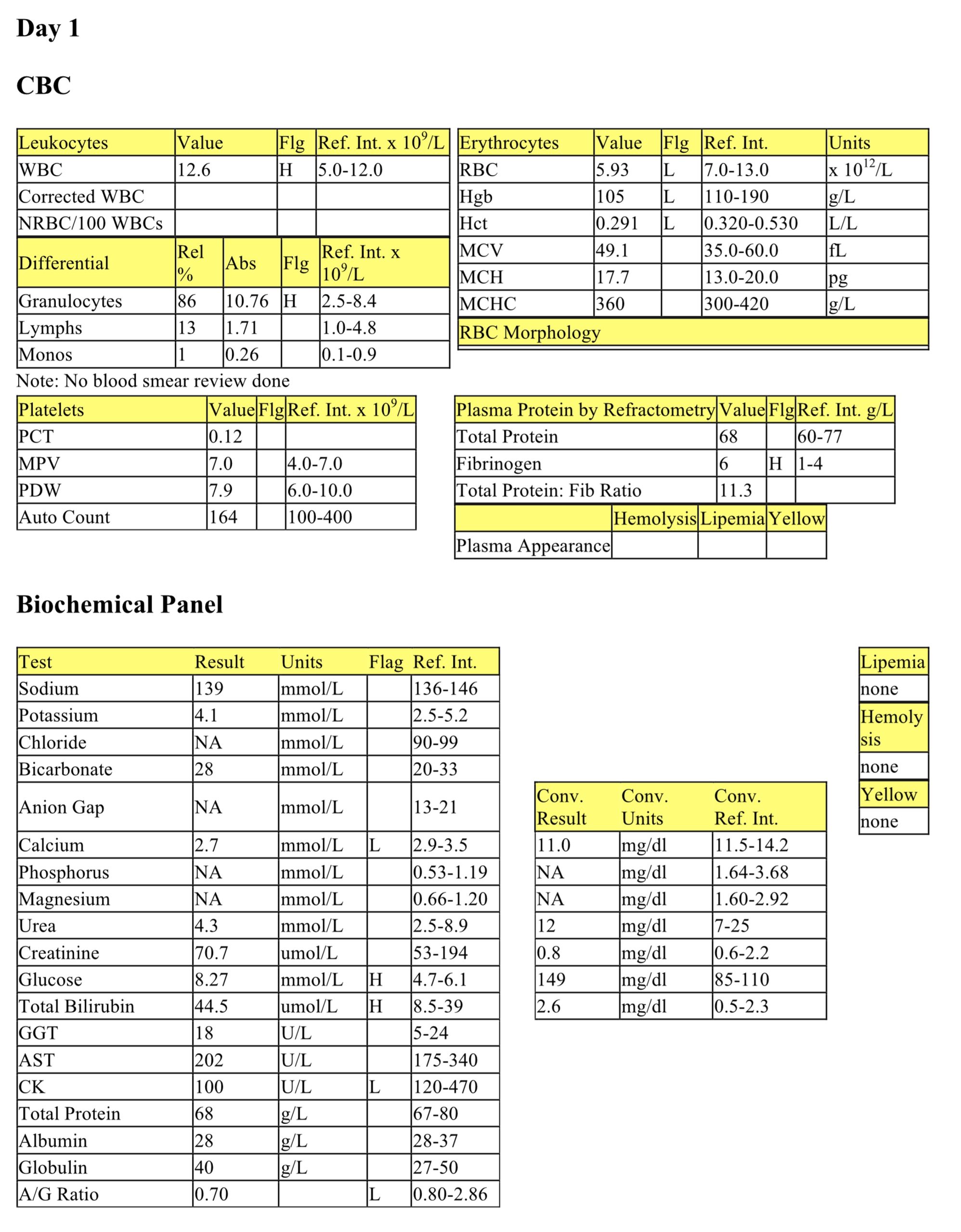
CBC day 1
There is a mild anemia. Given the species, regeneration cannot be assessed based on the CBC, however anemia of inflammatory disease is likely. There is a mild leukocytosis, characterized by a mild increase in granulocytes. This CBC was performed in-house, on an analyzer that does not differentiate the 3 types of granulocytes. Granulocytes are most likely neutrophils, although an eosinophilia cannot be ruled out without examining the blood smear. It is unknown if bands and toxic change are present, as there is no record of blood smear examination. Fibrinogen is increased without an increase in total protein; the total protein to fibrinogen ratio is <15, which is consistent with inflammation in the horse.
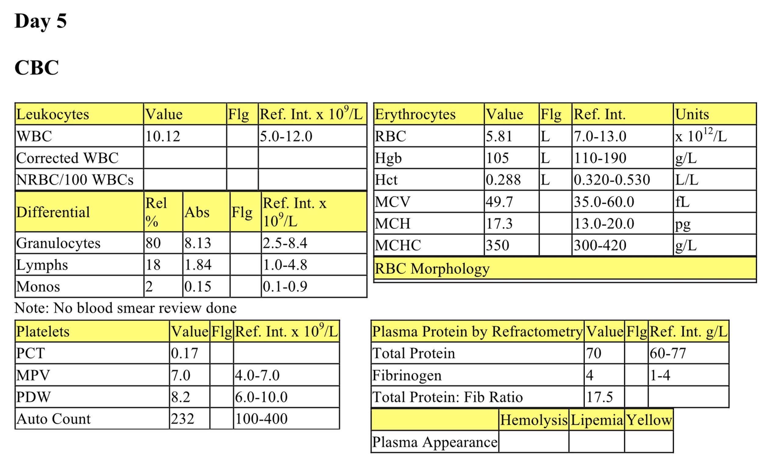
CBC day 5
The mild anemia persists. Leukocytes and granulocytes are now within RI, suggesting a resolution of inflammation. The fibrinogen concentration has decreased; the P:F ratio is now 17.5, reflecting improvement.
Biochemical Panel day 1
Mild hypocalcemia may relate to decreased intake and low normal albumin. Mild hyperglycemia likely reflects stress of illness. There is a mild hyperbilirubinemia which is likely secondary to anorexia. Mildly decreased CK activity is not clinically significant. Albumin concentration is low normal, however the A/G ratio is mildly decreased, suggesting albumin is decreased relative to globulins. Note that several parameters are not available (biochemical panel performed in house).
Trans-tracheal Wash
Degenerate neutrophils predominate in this sample, with low numbers of macrophages. Rare diplococci are present within neutrophils, with numerous mats of diplococci in the background. Small lakes of mucus and scant free erythrocytes and hematoidin crystals are seen. The wash reflects neutrophil-rich septic inflammation. Culture revealed Streptococcus sp and Actinobacillus equuli.
- Dolly had pleuropneumonia, likely precipitated by the stress of a long trailer ride from Texas. She was treated with appropriate antibiotics and recovered.
Case 2: Zed
Zed, a 19-year-old stallion of unknown breed, has a 6 week history of weight loss after being kicked and a 3 day history of right hind limb lameness.
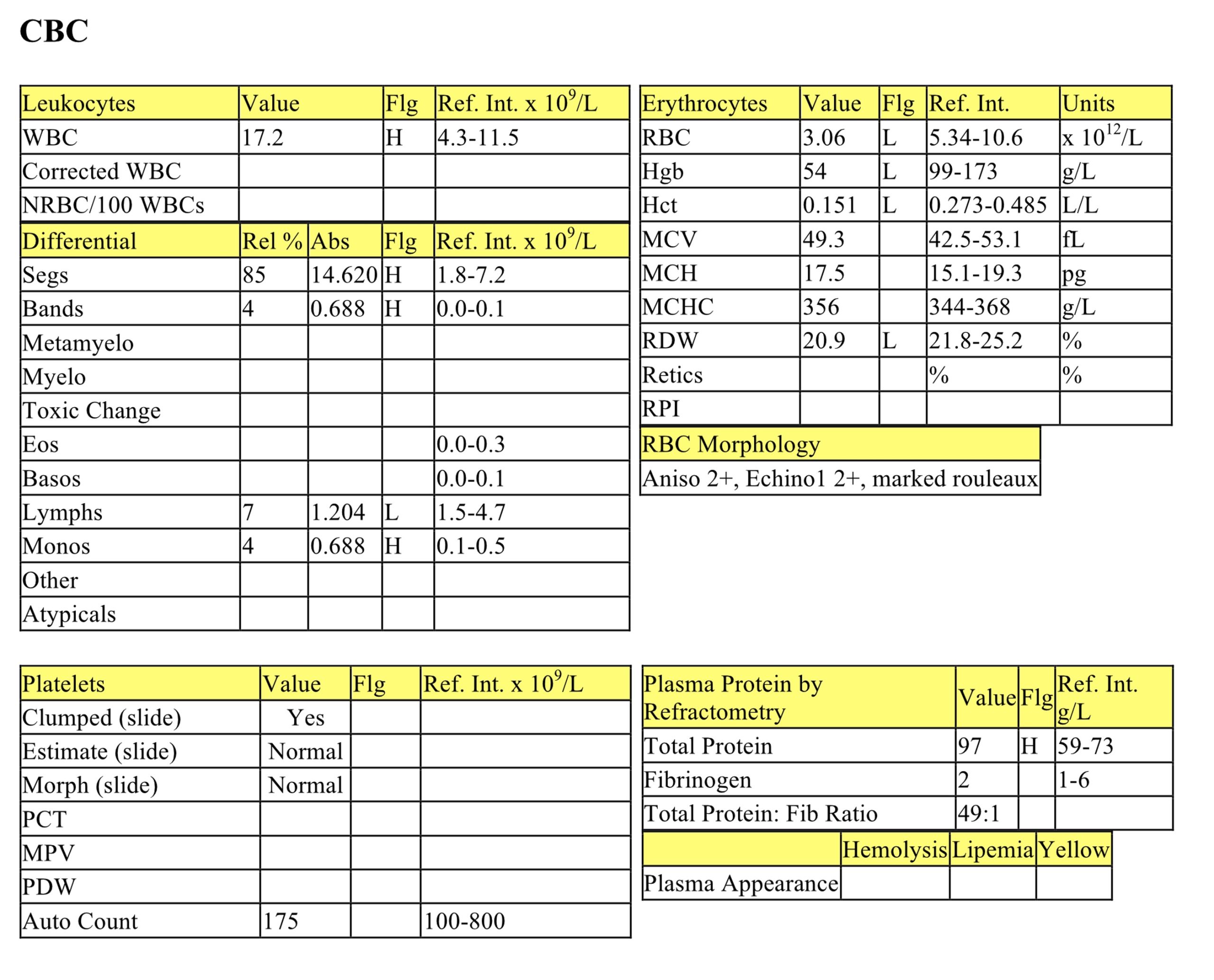
CBC
There is a moderate to marked anemia that is normocytic and normochromic. Since horses do not release reticulocytes, regeneration is difficult to assess on a single CBC. Regeneration in horses is supported by an increased MCV or the presence of macrocytes, however, neither of these is present in Zed’s CBC. A portion of the anemia may be attributed to inflammatory disease, however there could also be blood loss into the site of inflammation. Marked rouleaux are probably secondary to hyperproteinemia and hyperglobulinemia (see below). The leukogram shows a mild to moderate neutrophilia and a mild left shift and monocytosis consistent with inflammation. Mild lymphopenia reflects stress. See below for discussion of hyperproteinemia.
Biochemical Panel
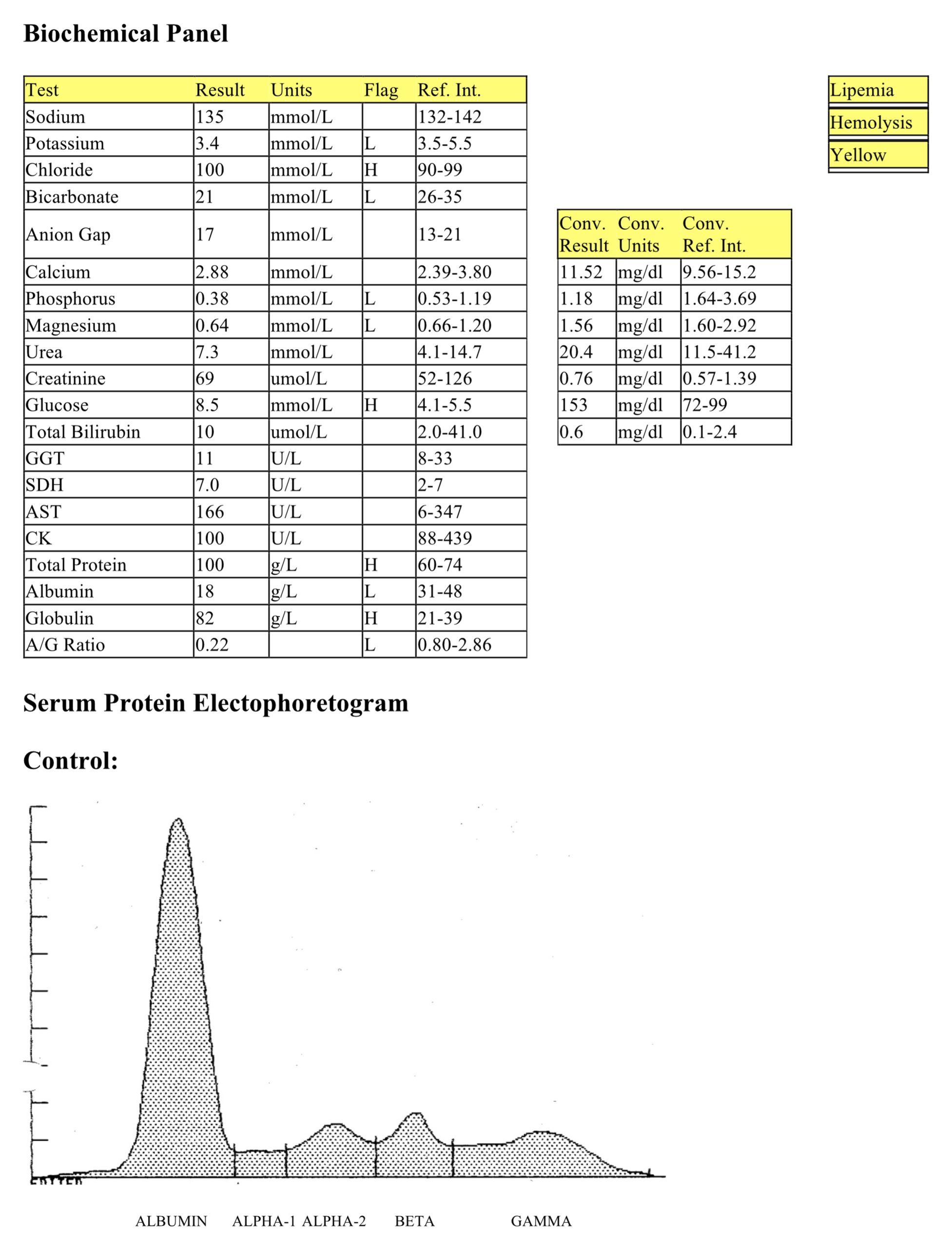
Mild hypokalemia may be due to decreased intake. Mild hyperchloremia (with low normal sodium) is likely secondary to metabolic acidosis given the mild decrease in bicarbonate. The normal anion gap suggests a loss of bicarbonate (via the gastrointestinal tract or kidneys) as the cause of the acidosis. Moderate hypophosphatemia and mild hypomagnesemia may be due to decreased intake. Hypomagnesemia could also be related to hypoalbuminemia and decreased protein binding. Mild hyperglycemia reflects stress. Marked hyperproteinemia due to hyperglobulinemia is likely due to chronic antigenic stimulation. Moderate hypoalbuminemia reflects decreased production (negative acute phase protein), and given the marked hyperglobulinemia, other causes of hypoalbuminemia such as those related to oncotic pressure should be considered.
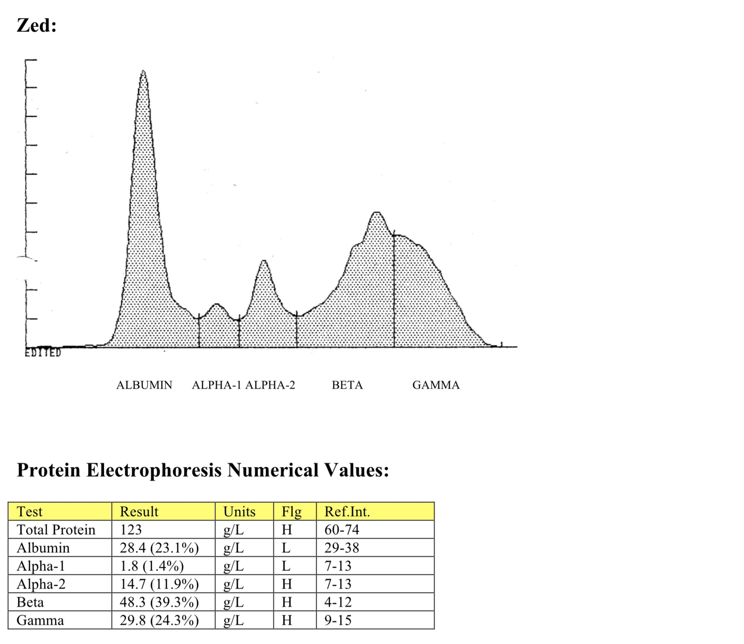
Serum Protein Electrophoresis
Hyperglobulinemia is due to an increase in alpha-2, beta, and gamma globulins. This can be determined by looking at the numerical results, however, the electrophoretogram is needed in order to determine the shape of the peak. The peak is broad based, consistent with chronic inflammation (polyclonal gammopathy).
- Aspiration of a fluid pocket on the lateral thigh revealed numerous degenerate neutrophils and intracellular cocci, reflecting septic inflammation. Staphylococcus aureus was cultured.
- Zed was treated with antibiotics and recovered, although secondary degenerative joint disease developed in the right stifle.
Additional Cases
Ch 1 Erythrocytes- Case 1 Odie
Ch 2 Leukocytes- Case 6 Tootsie
Ch 3 Hemopoietic Neoplasia- Case 2 Katie
Ch 7 Renal- Case 3 Marina, Case 4 Phillip
Ch 9 Digestive- Case 1 Pamela, Case 3 Sonia
Decrease in hematocrit (PCV) recognized on the complete blood count (CBC); usually hemoglobin concentration and RBC numbers are also decreased.
Mobile leukocyte with the ability to phagocytose, degrade, and/or kill microorganisms (neutrophil, eosinophil, basophil).
Increase in the number of eosinophils in peripheral blood.
Cytoplasmic abnormalities seen in neutrophils that have not matured normally in the bone marrow. Abnormalities include retention of primary granules, vacuolation, darker staining due to retention of ribosomes, and deposits of rough endoplasmic reticulum (Döhle bodies).
White blood cell (WBC); includes neutrophils, eosinophils, basophils, monocytes, lymphocytes, mast cells.
Term used to describe neutrophils in cytologic preparations indicating swelling and lysis of the neutrophil nucleus, e.g. due to bacterial infection.
Mononuclear phagocyte found in tissues that develops from circulating blood monocytes and fulfills many roles in normal immune function including antigen presentation.
Red blood cell (RBC); an anucleate (in mammalian species) cell containing hemoglobin needed for oxygen transport. Typically shaped like a bi-concave disk.
Granulocyte with fine, inconspicuous cytoplasmic granules and a segmented nucleus; important in phagocytosis and killing of bacteria.
Anucleate (in mammalian species), immature erythrocyte containing cytoplasmic RNA and ribosomes which are precipitated by staining with new methylene blue.
Average erythrocyte size in femtoliters (measured or calculated PCV or Hct÷RBC).
“Stack” of red blood cells resembling a roll of coins; prominent in equine and often feline blood. Formation increases as negative charges between RBCs decrease (e.g. with increased plasma proteins).
Increase in the number of neutrophils in peripheral blood.
Release of less mature neutrophil stages (bands, metamyelocytes, myelocytes) from the marrow into the peripheral blood in response to inflammation.
Increase in the number of monocytes in peripheral blood.
Difference between unmeasured anion and cation concentrations, calculated using the formula: (Na+ + K+) minus (Cl- + HCO3-.)
Gammopathy characterized by a broad-based peak in the gamma region and reflecting production of many different immunoglobulins from antigenic stimulation as may be seen with a chronic infection.

