Case Studies
Case 1. Shirley
Shirley, an adult Holstein-Friesian cow, was anorexic since calving the previous day. She had been treated for milk fever but was still not eating.
CBC
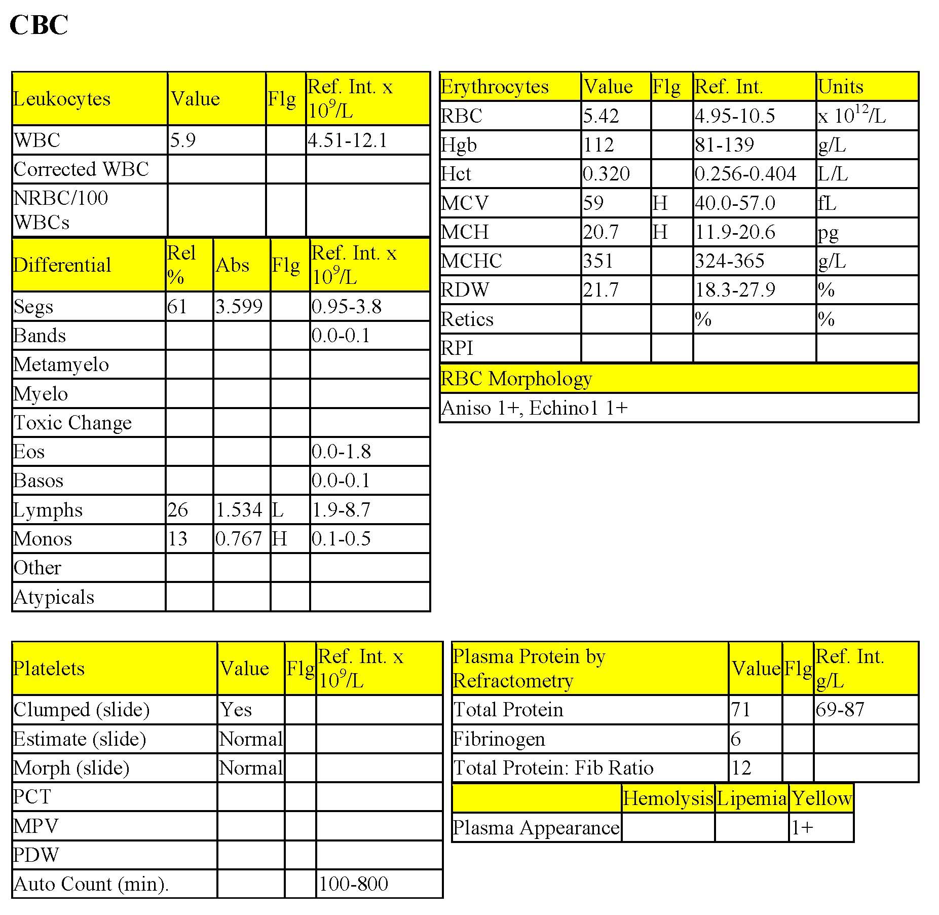
The erythrogram is unremarkable. The mild elevations in MCV and MCH, in isolation are unlikely to be clinically significant. Total WBC count is within the RI. Neutrophil numbers are high normal and there is a mild lymphopenia and mild monocytosis. These findings are consistent with stress (high levels of endogenous cortisol due to illness). The fibrinogen is within the RI, which is consistent with the lack of evidence of inflammation on the leukogram. The total protein to fibrinogen ratio is not evaluated unless the fibrinogen is elevated, which is not the case here. Yellow plasma may be seen in cattle due to -carotenes in grass and hay; however, it can also signal hyperbilirubinemia (see biochemical panel interpretation).
Biochemical Panel
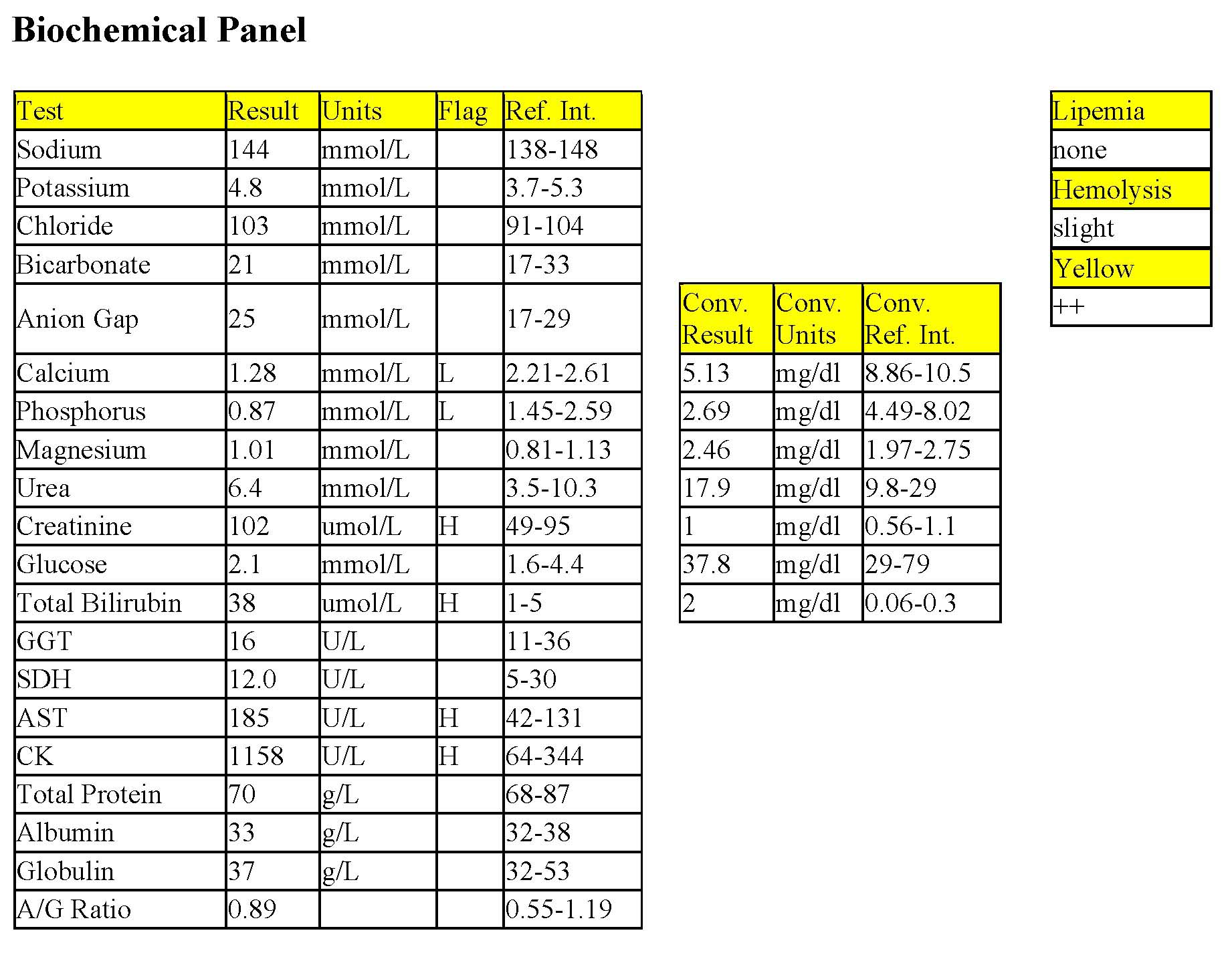
There is a moderate to severe hypocalcemia and moderate hypophosphatemia, consistent with the diagnosis of milk fever (despite prior treatment for this condition). Mildly increased creatinine suggests reduced renal perfusion. Urea is not concurrently elevated possibly due to low protein in the diet, excretion of urea through the digestive tract where it serves as a source of protein in ruminants, or both. Mild to moderate hyperbilirubinemia may relate to anorexia resulting in competition between bilirubin and mobilized fatty acids for uptake by hepatocytes and impaired hepatic blood flow resulting in decreased uptake of unconjugated bilirubin, decreased conjugation of bilirubin, or both. CK and AST activities are mildly elevated possibly due to myocyte leakage from increased recumbency associated with hypocalcemia, although this was not provided in the history.
- Despite receiving treatment for milk fever, Shirley is still hypocalcemic; clinical signs are expected at calcium concentrations <1.6 mmol/L. Cattle normally have about twice as many lymphocytes as neutrophils. A reversal in this ratio is seen with stress in the cow, and this is present in this case. Even high normal neutrophil numbers and low normal lymphocyte numbers (without any flags for values outside of the RI) would represent a stress response. This emphasizes the importance of looking at all data, not only those that are flagged.
Postparturient cattle can develop serious inflammatory as well as metabolic diseases. The CBC was important in determining that Shirley did not have inflammatory disease. No further follow up is available.
Case 2. Socks
Socks, a 9-year-old M(c) DLH cat, had a history of vomiting, anorexia, and pyrexia.
CBC
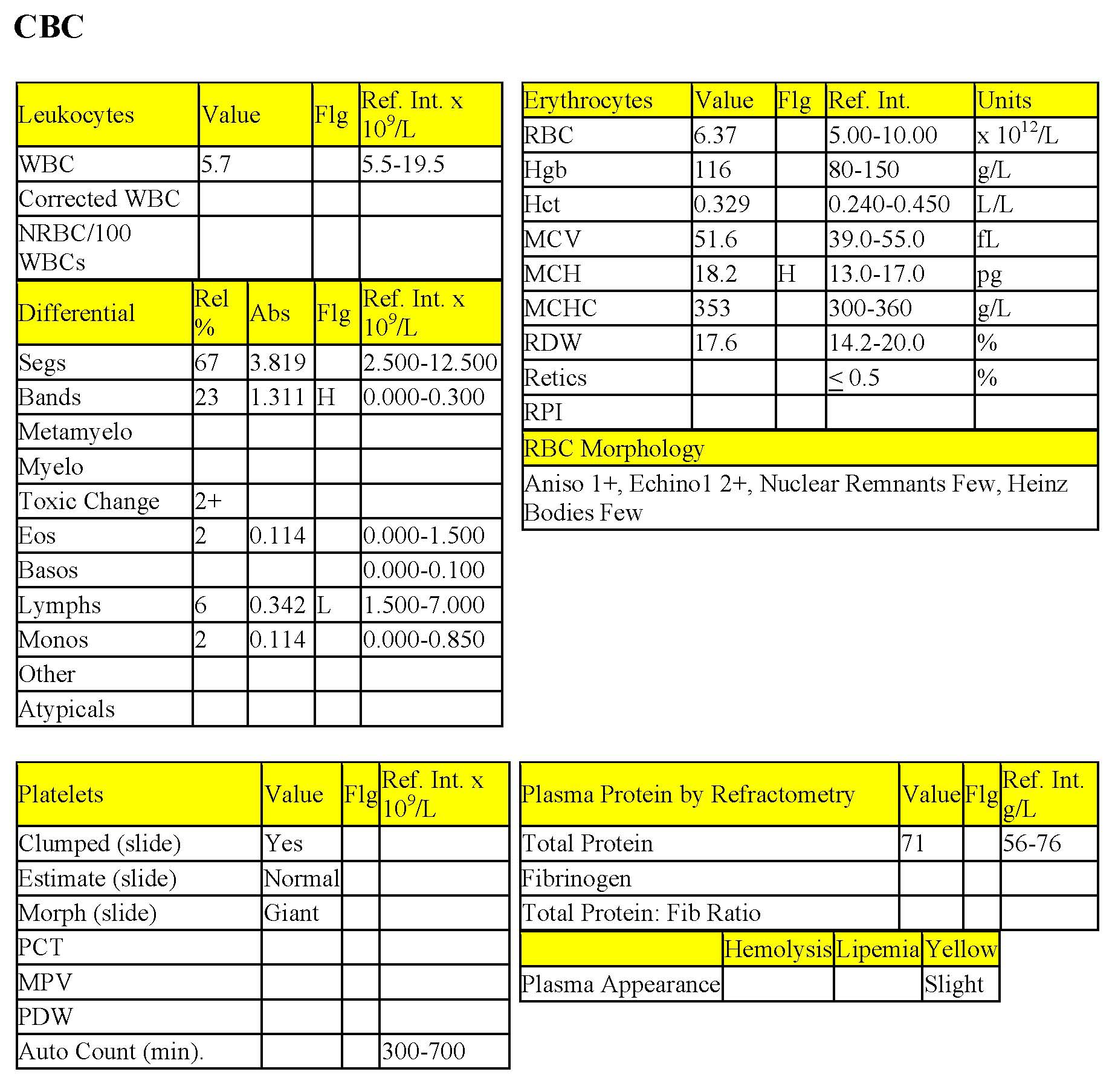
The erythrogram is unremarkable. The mildly increased MCH is not clinically significant. Heinz bodies are often seen in cats and are not considered to be significant when small, low in number, and not accompanied by hemolytic anemia (see Chapter 1: Erythrocytes). The total WBC count is low normal. Although segmented neutrophils are within the RI, they are low normal and there is a mild to moderate left shift with moderate toxic change, and severe lymphopenia. These findings are consistent with acute inflammation and stress. This leukogram could be described as “in transition”, and a follow-up CBC in 24-36 hours would help to determine if the bone marrow is able to meet the demand for neutrophils. The toxic change indicates accelerated release of neutrophils from the bone marrow and a lack of maturation of the neutrophil cytoplasm. Total protein, by refractometry, is within RI, and Socks does not seem to be dehydrated based on the CBC findings. Serum biochemistry was not done.
- Socks was diagnosed with gastroenteritis and bone fragments were detected on imaging studies. Surgery was recommended but not done. Socks did not respond to conservative therapy and was later euthanized. Neither follow-up CBC nor necropsy was done.
Case 3. Ranger
Ranger, a M DSH cat of unknown age, had a laceration under the neck for 3 days. He was presented in a collapsed state and was hypoglycemic on glucometer reading.
CBC
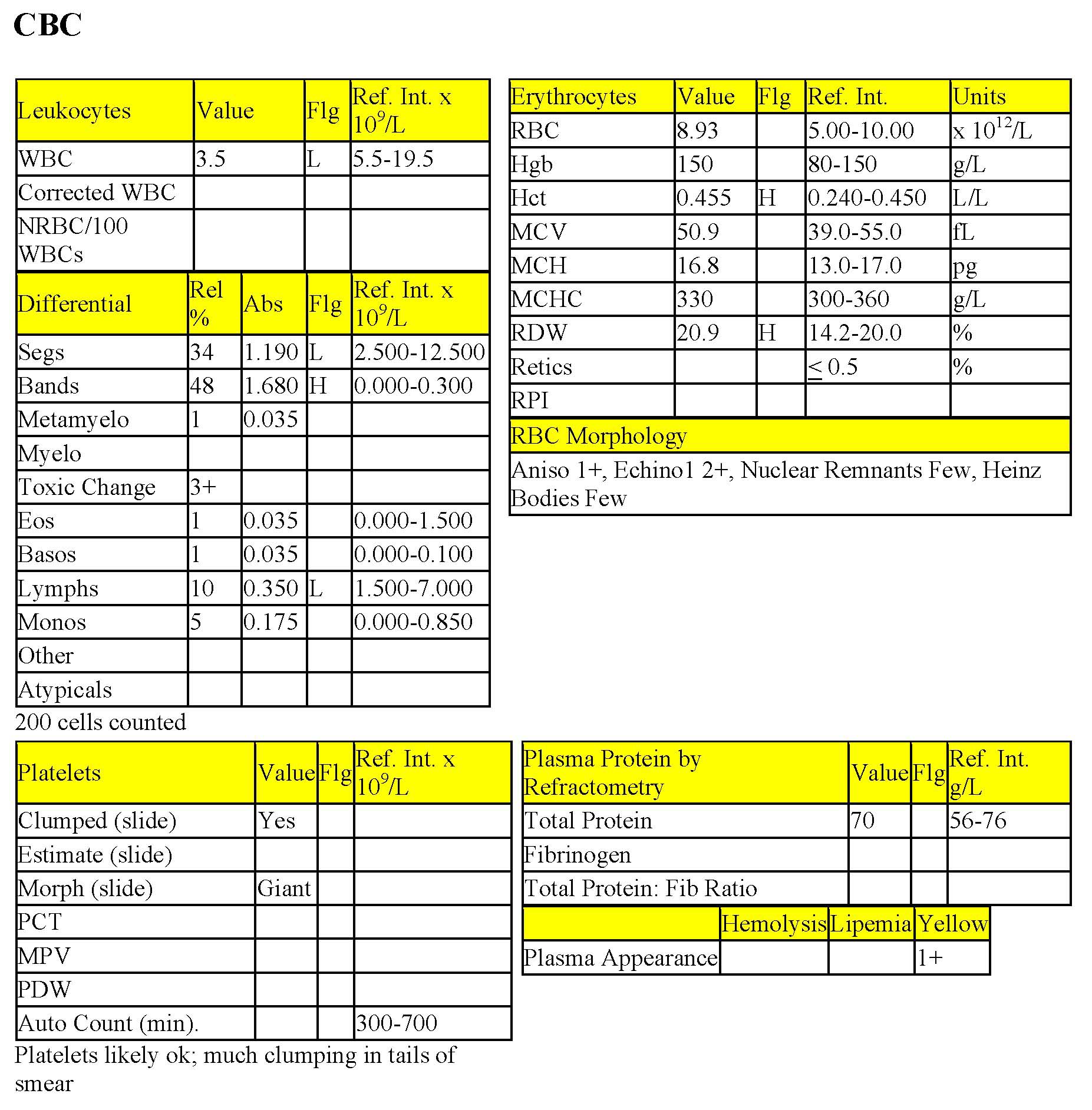
The Hct is borderline high which could indicate dehydration, although this is not reflected in the total protein measured by refractometry. There is a moderate leukopenia which is characterized by moderate neutropenia, moderate left shift (with both bands and metamyelocytes being released), severe toxic change, and severe lymphopenia. This leukogram represents a degenerative left shift (see Neutropenia and Pearls sections) and indicates acute, overwhelming inflammation, and stress. Ranger’s bone marrow has been depleted of its post-mitotic storage pool due to the severity of the peripheral demand for neutrophils. Yellow plasma suggests hyperbilirubinemia, although this would have to be confirmed on serum biochemistry. Hyperbilirubinemia can be seen with severe inflammatory conditions associated with endotoxemia (see Chapter 8. Hepatobiliary System). The prognosis is poor unless the marrow has an opportunity to respond through effective treatment of the focus of inflammation, likely to be related to an infected neck laceration in this patient. Hypoglycemia can be seen with endotoxemia, which is supported by other findings in this case.
- Based on the clinical and laboratory findings, and poor prognosis, Ranger was euthanized. A necropsy was not done.
Case 4. Harriet
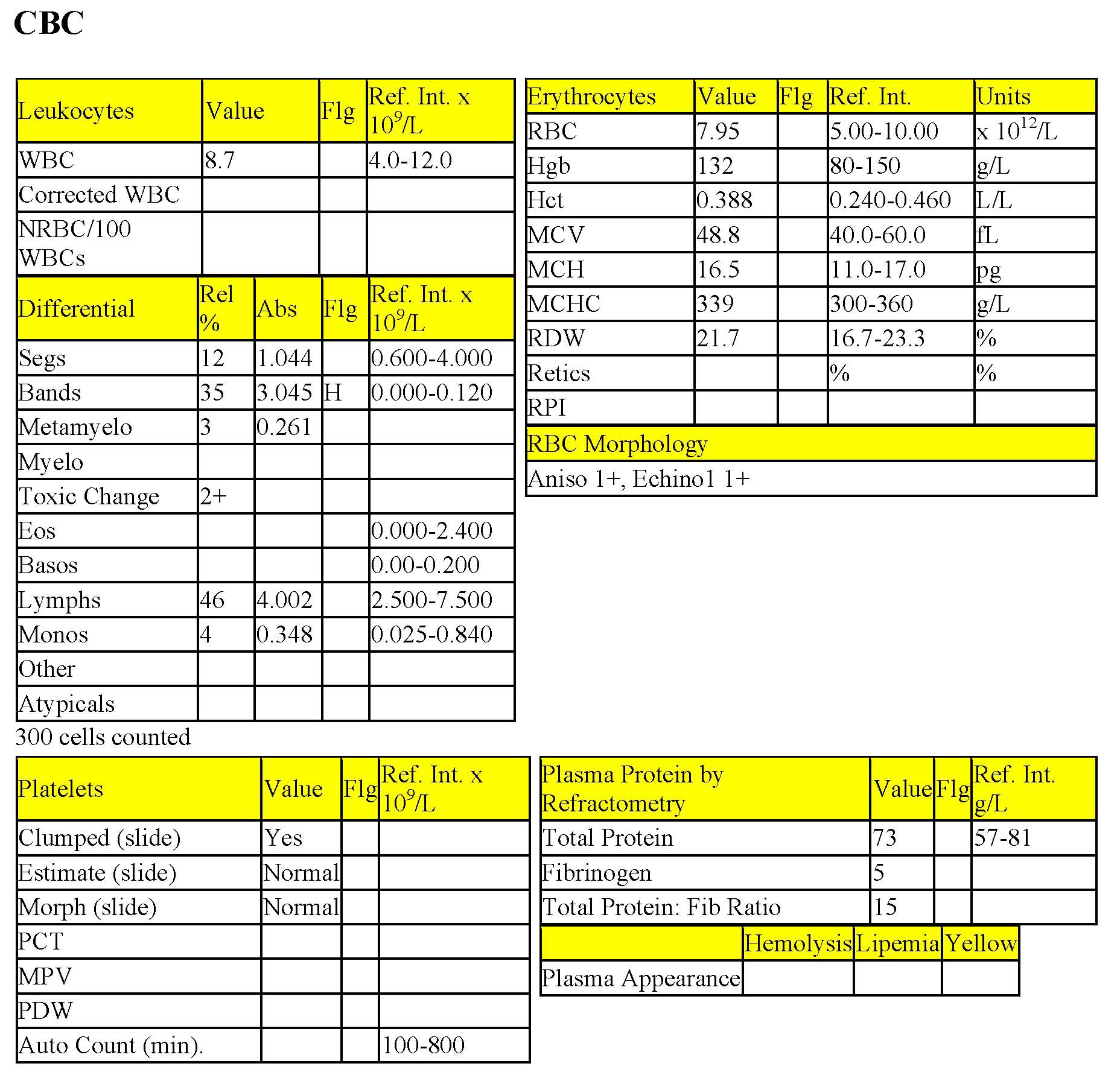
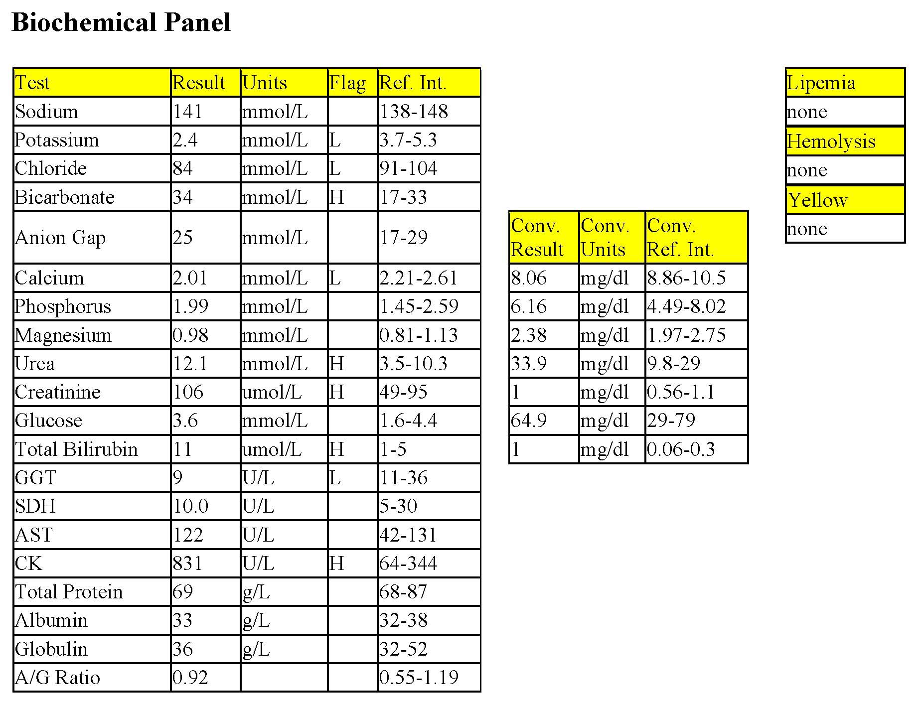
Harriet, a 2-year-old Red Angus cow, had a history of lethargy since calving 5 days earlier.
CBC
The erythrogram is unremarkable. Total WBC count is within the RI; however, segmented neutrophils are low normal and there is a moderate left shift with moderate toxic change. This leukogram represents a degenerative left shift and indicates acute, severe inflammation. Despite this finding, there is no concurrent stress lymphopenia or hyperfibrinogenemia.
Biochemical Panel
Moderate hypokalemia suggests decreased intake, increased loss (renal or intestinal), and/or redistribution from the extracellular to the intracellular space due to the metabolic alkalosis. Mild hypochloremic metabolic alkalosis may be due to inappetence and abomasal stasis leading to pooling of HCl. Mild hypocalcemia could be explained by decreased intake and is not sufficiently low to suggest milk fever. Mild azotemia should be interpreted in relation to hydration status and urine concentrating ability (urinalysis not available). Mild hyperbilirubinemia is common in cattle that are off feed, and is explained by decreased hepatic uptake and conjugation of bilirubin. There is no pathologic significance to the decreased GGT. CK activity is mildly increased which could be related to muscle injury secondary to recent calving, increased recumbency, or both.
- Harriet has an inflammatory focus. Common sites in postparturient cattle are: mammary gland, endometrium, and peritoneal cavity. On rectal examination, peritonitis was suspected. Neither a repeat CBC nor abdominocentesis was done and no follow-up information is available.
Case 5. Sunny
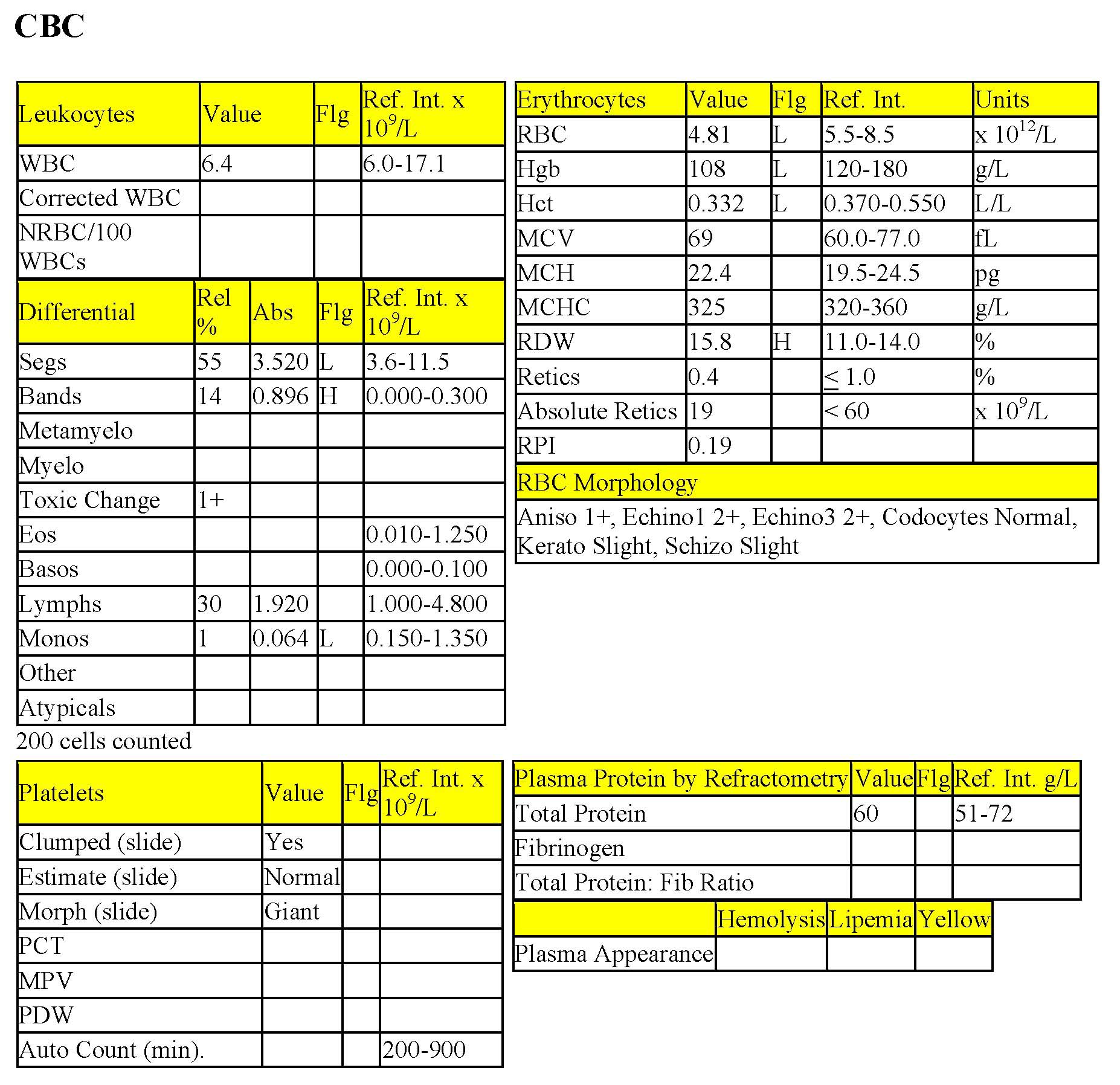
Sunny, a 2-month-old M Great Pyrenees puppy, had diarrhea and inappetence for 2 days.
Sunny, day 1
CBC (day 1)
There is a mild nonregenerative anemia which may be partially age-related or due to recent blood loss, or a combination of the two. If Sunny is dehydrated, the degree of anemia may be underestimated. Although the RDW is mildly increased, this does not correspond to an increase in anisocytosis on smear evaluation and may not be significant. Echinocytes III can be associated with electrolyte and acid-base disturbances and azotemia; in vitro formation can also occur. Keratocytes and schizocytes are only slight, but can be seen with fibrin strand injury, such as with disseminated intravascular coagulation and fibrinolysis.
Total WBC count is low normal. There is a very mild neutropenia with a mild left shift, mild toxic change, and mild monocytopenia. The lack of stress lymphopenia could relate to antigenic stimulation/recent vaccination in this puppy. Total protein, by refractometry is within the RI, but should be interpreted with regard to hydration status and possible protein loss via the GI tract. Also, protein is better assessed by serum biochemical analysis.
CBC (day 3)
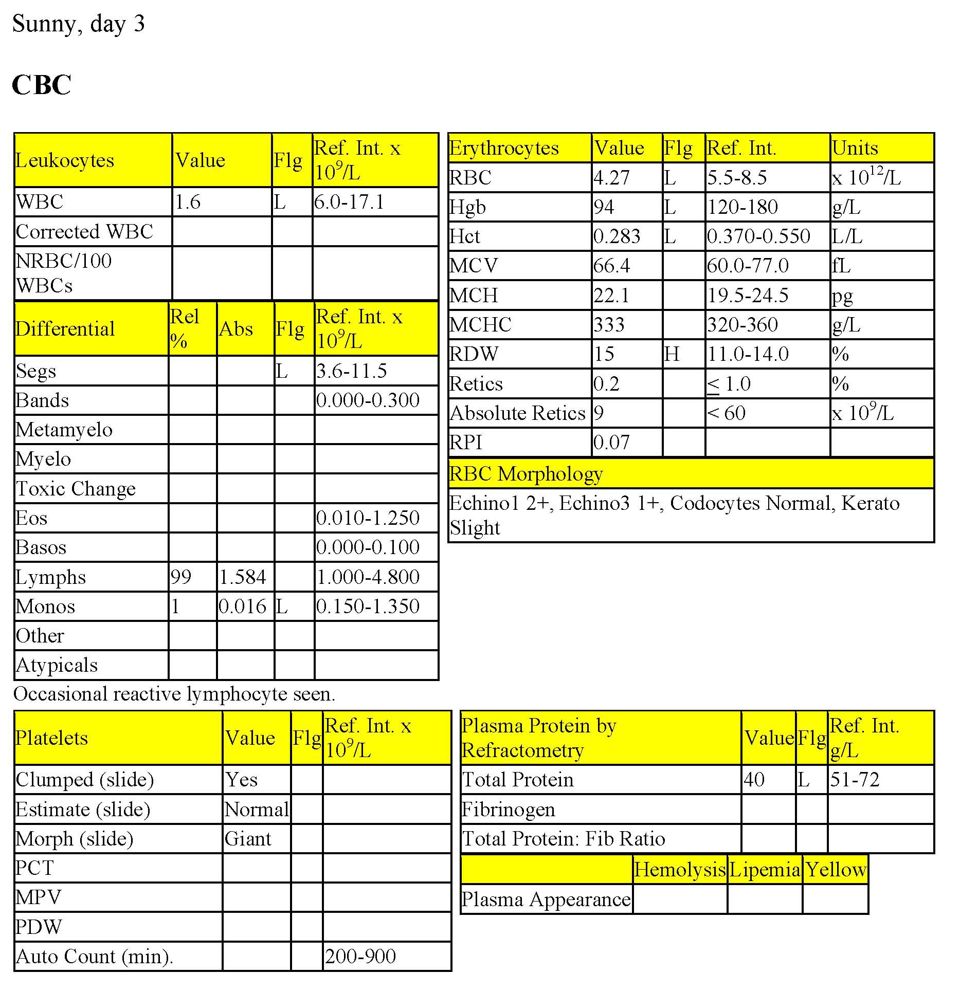
The Hct is slightly lower and hypoproteinemia is now present – these changes may relate to the dilutional effect of fluid therapy, blood loss, or both. There is a marked leukopenia which is due to severe neutropenia (no neutrophils seen) and moderate monocytopenia. The lack of stress lymphopenia and presence of reactive lymphocytes could relate to antigenic stimulation/recent vaccination.
These findings are highly suggestive of canine parvoviral infection. An ELISA to detect parvovirus antigen in feces was positive.
- Sunny started to vomit and he developed hemorrhagic diarrhea. He was euthanized based on his deteriorating clinical condition and worsening leukogram. The second CBC helped with prognostication since it reinforced the clinical impression that Sunny was becoming sicker in spite of appropriate therapy.
- Note: The leukogram in this case demonstrates how deceptive the leukocyte % can be. Notice that on the day 3 differential, 99% of the leukocytes are lymphocytes, compared to 30% on day 1. However the absolute counts are very similar and are bordering on low normal.
Case 6. Tootsie
Tootsie, a mature mixed breed beef cow, had fever and weight loss.
CBC
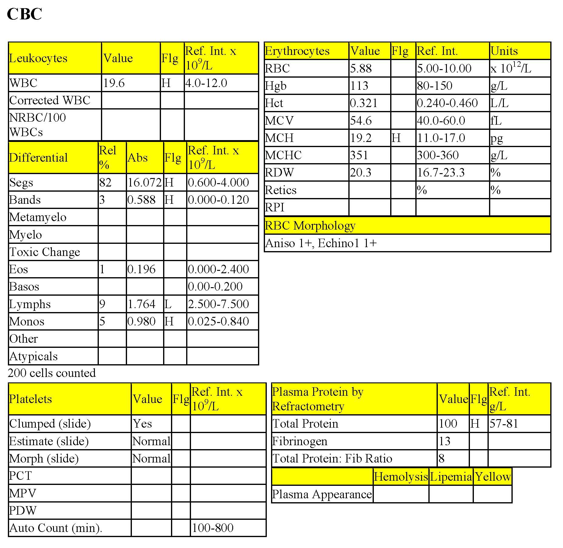
The erythrogram is unremarkable. The mildly increased MCH is unlikely to be clinically significant. There is a moderate leukocytosis characterized by a moderate neutrophilia with a mild left shift and mild monocytosis. There is also a mild lymphopenia. Total protein is moderately elevated. There is a moderate to marked hyperfibrinogenemia which is absolute, based on the total protein to fibrinogen ratio of <10. These findings are consistent with chronic inflammation and stress.
Biochemical Panel
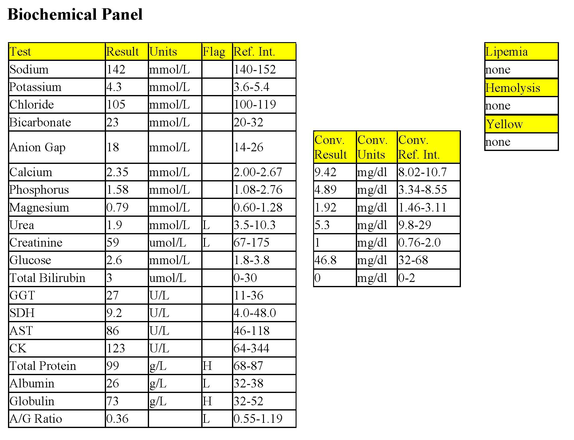
The low urea could be due to low protein intake. Hepatic dysfunction is an unlikely cause of the low urea given the lack of changes in other indicators of hepatic failure. Decreased creatinine is probably from reduced muscle mass. The elevated total protein, low albumin, high globulins, and low albumin to globulin ratio, can be explained in several ways. Hyperglobulinemia could be seen with chronic antigenic stimulation; also, increases in acute phase proteins associated with inflammation could increase globulins relative to albumin. Albumin is a negative acute phase protein and its synthesis is decreased with inflammation. Albumin could also be low from low protein intake, if prolonged, or increased losses through the kidney or intestinal tract (although with intestinal pathology leading to hypoproteinemia, both hypoalbuminemia and hypoglobulinemia would normally be expected).
Although the leukogram changes do not seem dramatic relative to other species with chronic inflammation, cattle have a smaller pool of proliferating and maturing neutrophils. Bone marrow examination would be expected to reveal neutrophilic hyperplasia in this case. It was somewhat surprising that Tootsie had not developed a nonregenerative anemia due to inflammatory disease; however, possibly her normal hematocrit is closer to the high end of the RI.
- It was later learned that Tootsie had been treated with antibiotics for several days and that she had clinical evidence of unresponsive respiratory disease.
Case 7. Hailey
Hailey, a 9-year-old F(s) Cocker Spaniel dog, had lethargy and inappetence.
CBC
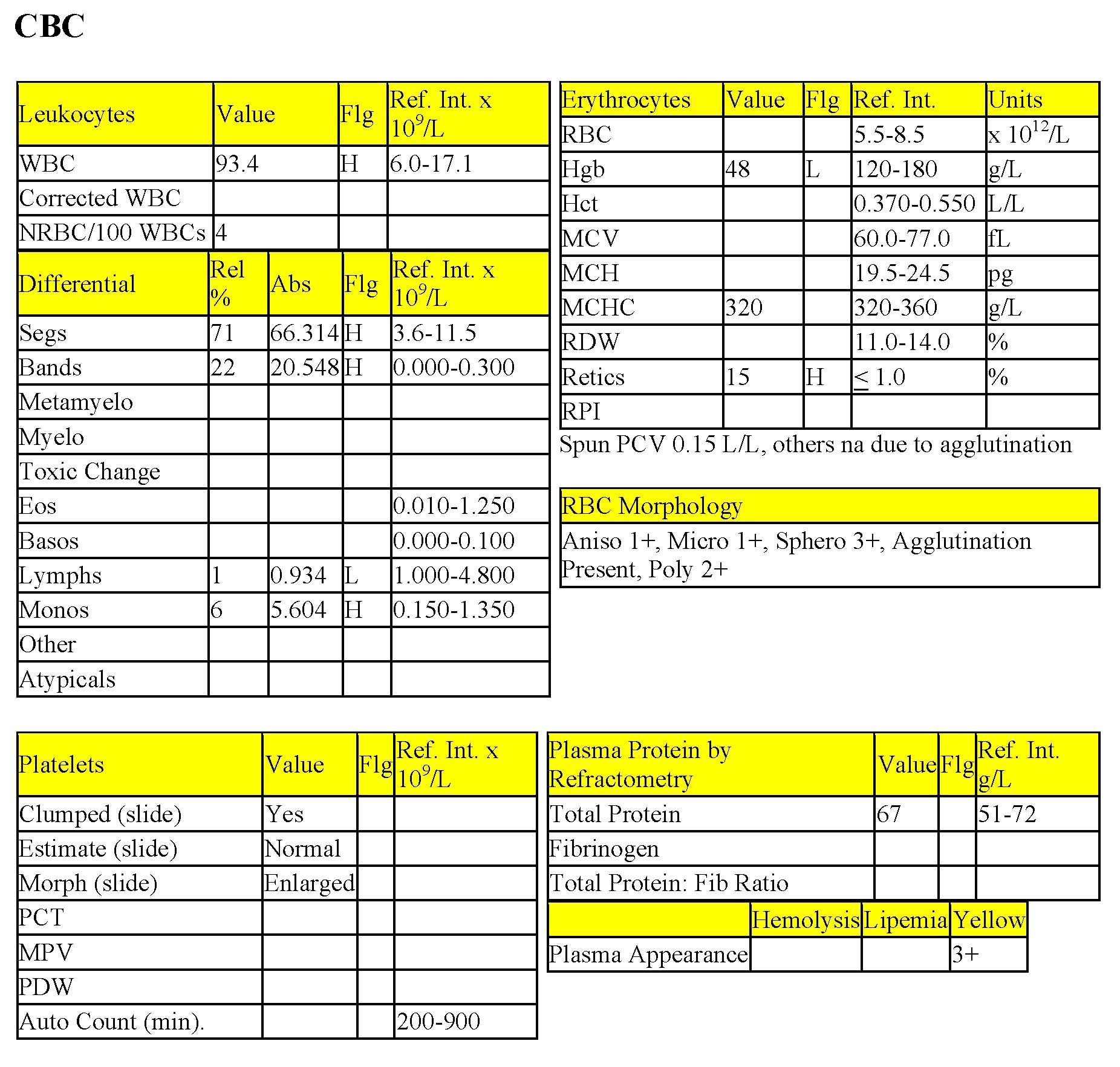
Hailey has a severe anemia with marked regeneration. The absolute reticulocyte count cannot be calculated without a RBC count, which is unavailable due to RBC agglutination. Agglutination and spherocytes noted on smear evaluation are consistent with immune-mediated hemolysis. Many erythrocyte measurements and calculations could not be made due to the agglutination. Yellow plasma (icterus) is consistent with extravascular hemolysis in this case. The severe leukocytosis is characterized by severe neutrophilia, severe left shift, and moderate monocytosis. These changes would normally suggest severe chronic inflammation; however, the history with IMHA usually does not suggest chronicity. A possible explanation is that the inflammatory cells are remaining within the circulation due to the RBC destruction that is occurring and are not migrating into tissues. Lymphopenia is consistent with stress.
- Immune-mediated hemolytic anemia may by accompanied by an inflammatory leukogram of this magnitude. The dramatic changes that may be seen on the leukogram may divert attention from the primary problem, which is the destruction of RBCs. However, moderate to marked leukocytosis may also signal concurrent tissue injury due to hypoxia, thromboembolic disease, or both.
Case 8. Jimmy
Jimmy, a young adult M(c) DSH cat, had extreme lethargy and weakness.
CBC
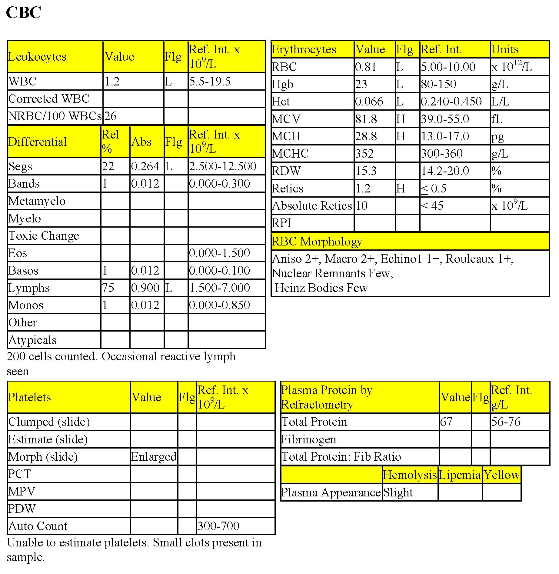
Jimmy has a severe anemia which is nonregenerative, but macrocytic (high MCV with corresponding high MCH, and macrocytes noted on RBC morphology). Despite the lack of regeneration (reticulocytes are not increased and no polychromasia noted), nucleated RBCs are present. There is a severe leukopenia characterized by severe neutropenia, without a significant left shift, and mild lymphopenia (panleukopenia).
The bicytopenia may indicate a bone marrow disorder. Macrocytosis of RBCs, in the presence of nonregenerative anemia, is suggestive of asynchronous maturation of the nucleus relative to the cytoplasm (also called megaloblastic change). The cytoplasm matures normally, but the nucleus does not. RBC cytoplasms become hemoglobinated while the nucleus remains immature. Sometimes this condition can be seen in peripheral blood nucleated RBCs, otherwise, bone marrow examination may be necessary. Bone marrow examination would also be necessary to determine whether the cytopenias were due to a degenerative condition, maturation disturbance, or neoplastic process. These findings are most commonly associated with the feline leukemia virus (FeLV) and testing for viral antigen in the serum by ELISA, viral antigen in tissues by immunohistochemistry, and/or viral DNA in blood or tissues using the polymerase chain reaction (PCR) test would be indicated.
- Jimmy was tested for FeLV antigen and feline immunodeficiency virus (FIV) antibody by ELISA and was positive for FeLV. Jimmy was euthanized and a necropsy was not performed.
All tests on the complete blood count (CBC) that evaluate erythrocytes, including morphologic examination on the peripheral blood smear.
Average erythrocyte size in femtoliters (measured or calculated PCV or Hct÷RBC).
Average amount of hemoglobin per erythrocyte (calculated: hemoglobin÷RBC count).
Break-down product of hemoglobin.
Anemia due to destruction of RBCs. Lysis may be intravascular (within blood vessels) or extravascular (within tissues rich in mononuclear phagocytes such as spleen and liver), or both.
Fully mature neutrophil with a lobulated (segmented) nucleus.
Method of measuring the protein content of a fluid that relies on refraction of light, which is proportional to the quantity of solids in solution.
Process that adds base (HCO3-) to the blood or removes acid (H+); blood pH may or may not be increased.
Increases serum urea and/or creatinine.
Process of obtaining abdominal fluid for cytologic evaluation.
Variation in cell size.
Erythrocyte with even projections all around its membrane; often an artifact but can be a pathologic change. Echinocytes are classified as types 1 and 3 depending on the length and sharpness of the projections (spicules).
Crescent-shaped cells that are formed from mechanical shearing (usually due to fibrin strand deposition) of the red cell.
An erythrocyte fragment created by shearing trauma to erythrocytes within the vascular system; often the fragment which has been sheared off to form a keratocyte.
See Secondary hemostasis.
Process in which fibrin is broken down, resulting in clot dissolution.
General term for a decrease in number of any cell line in peripheral blood.
Decrease in the number of monocytes in peripheral blood.
Most abundant plasma protein in health; maintains oncotic pressure.
Varied group of plasma proteins including acute phase proteins and immunoglobulins.
Volume of erythrocytes per liter of whole blood. Reported as L/L (calculated: MCV x RBC count). Equivalent to PCV (%) determined by centrifugation of blood in a microhematocrit tube
Number of erythrocytes per liter of whole blood (measured).
Clumping of erythrocytes due to antibody interactions. Must be differentiated from rouleaux formation.
Erythrocyte with a spherical rather than biconcave disk shape, which results in a compact cell with a lack of central pallor. Often associated with immune-mediated hemolytic anemia.
Jaundice; deposition of pigment in skin, mucous membranes and sclera due to hyperbilirubinemia; usually results in a yellow discoloration of skin and mucous membranes.
Erythrocyte destruction within macrophages of the spleen, liver, lymph nodes, and bone marrow; often accompanied by hyperbilirubinemia and bilirubinuria.
Anemia due to destruction of RBCs that is caused by antibodies produced by the animal’s own immune system. Usually highly regenerative, with presence of spherocytes and agglu
Anucleate (in mammalian species), immature erythrocyte containing cytoplasmic RNA and ribosomes which are precipitated by staining with new methylene blue.
Increased numbers of anucleate, immature erythrocytes that stain bluish-pink with Romanowsky stains due to the presence of cytoplasmic RNA.
Increased number of erythrocytes with increased volume that usually corresponds with increased MCV; may be seen in regenerative anemias and can be normal in Poodle dogs.
Technique by which cells in tissue (e.g. poorly differentiated neoplastic cells) can be identified based on cell surface markers.

