Exudates
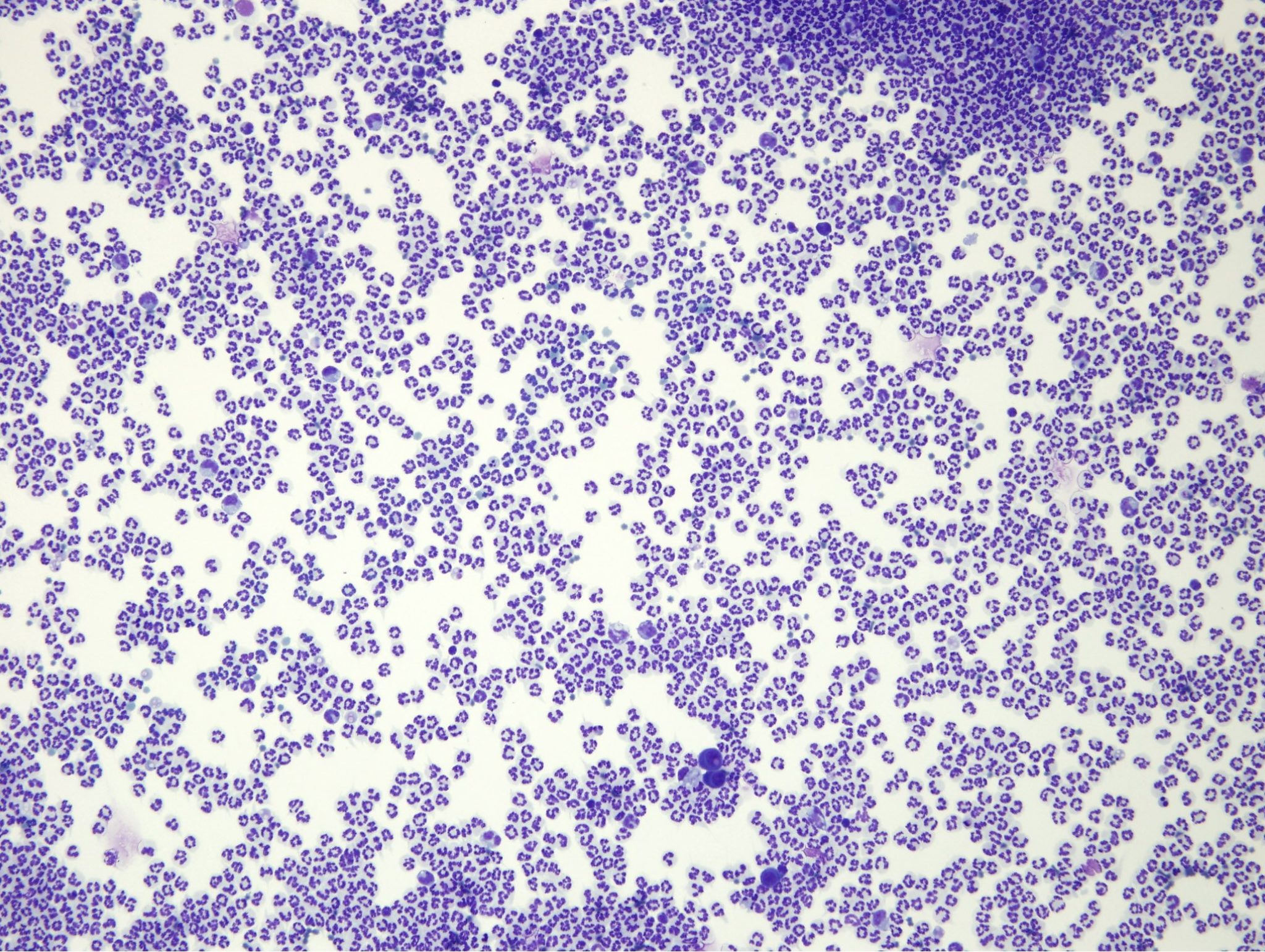
Exudates form when there is increased vascular permeability and demand for inflammatory cells, resulting in a fluid with an elevated protein concentration and nucleated cell count (protein >30 g/L and nucleated cell count >7 x 10⁹/L). Exudates may be septic or nonseptic; however, septic exudates produce the most dramatic elevations in nucleated cell count. Nonseptic exudates usually contain nondegenerate neutrophils, macrophages, mesothelial cells, and possibly low numbers of lymphocytes, plasma cells, and mast cells (Fig. 5.33). This type of fluid can result from a long-standing modified transudate, neoplasm, nonmicrobial irritant such as foreign material, bile, chyle (long-standing) or seminal fluid, and feline infectious peritonitis (FIP) virus.

Septic exudates often contain degenerate neutrophils, and the causative agent may be identified on microscopic examination (Fig. 5.8; see also Case 2). Highly pathogenic bacteria are more likely to cause degenerate change in neutrophils, particularly those which have phagocytosed bacteria.
Occasionally, inflammatory processes produce fluids which, according to protein concentration and nucleated cell count, would be classified as modified transudates, emphasizing the limitations of these guidelines in certain cases. For example, the bone marrow may be depleted of inflammatory cells (particularly neutrophils) with acute, overwhelming inflammation, producing a fluid with a high protein concentration unaccompanied by a high nucleated cell count. In this situation, the leukogram would be characterized by a degenerative left shift, and band and metamyelocyte neutrophils may even appear, not just in the peripheral blood but also in the fluid. Also, if the inflammatory process is marked by high protein fluid effusion, with a proportionately lower cellular response, the fluid may not fall within the exudate category. FIP virus often causes this type of effusion, though clearly, inflammation (due to immune complex vasculitis) is the pathologic event.
Exudate containing an infectious/etiologic agent (e.g. bacteria); often containing degenerate neutrophils as well.
Exudate without a causative agent, not resulting from infection.
All tests on the CBC that evaluate leukocytes. Also, that part of the leukon which is evaluated by examination of a peripheral blood sample (typically does not include leukocyte precursors).
Increase in immature neutrophils in which there is no neutrophilia and numbers of mature neutrophils are equal to or less than the numbers of immature stages; suggests the bone marrow is not able to meet peripheral demand.
Stage in granulopoiesis following myelocyte; nucleus begins to become indented.

