Chapter 3: Hemopoietic Neoplasia
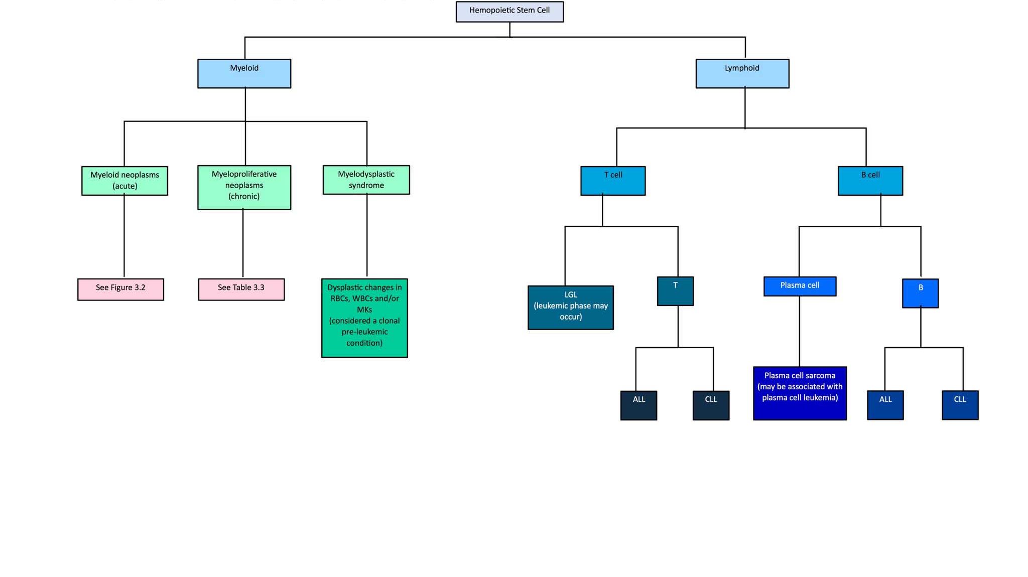
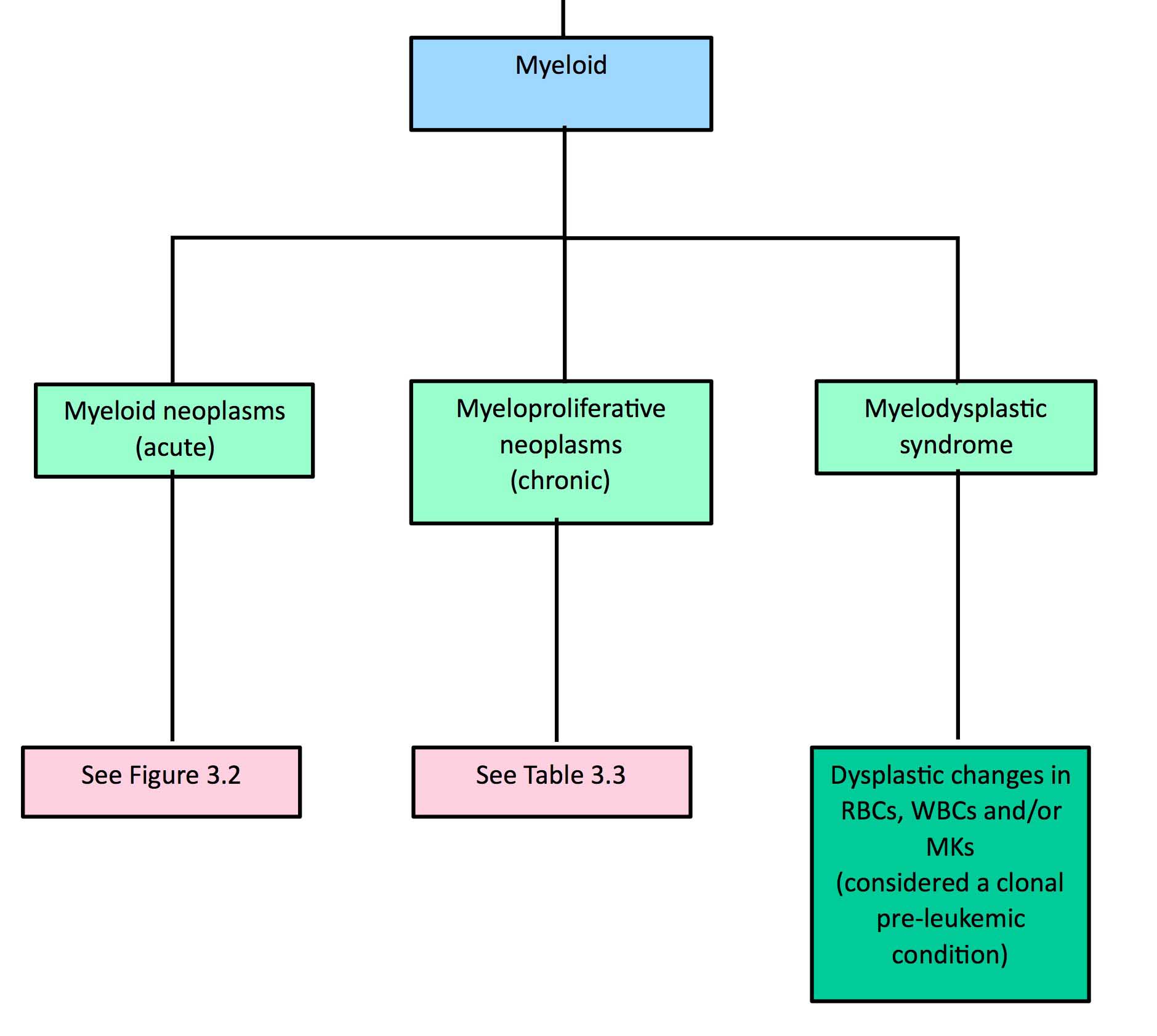
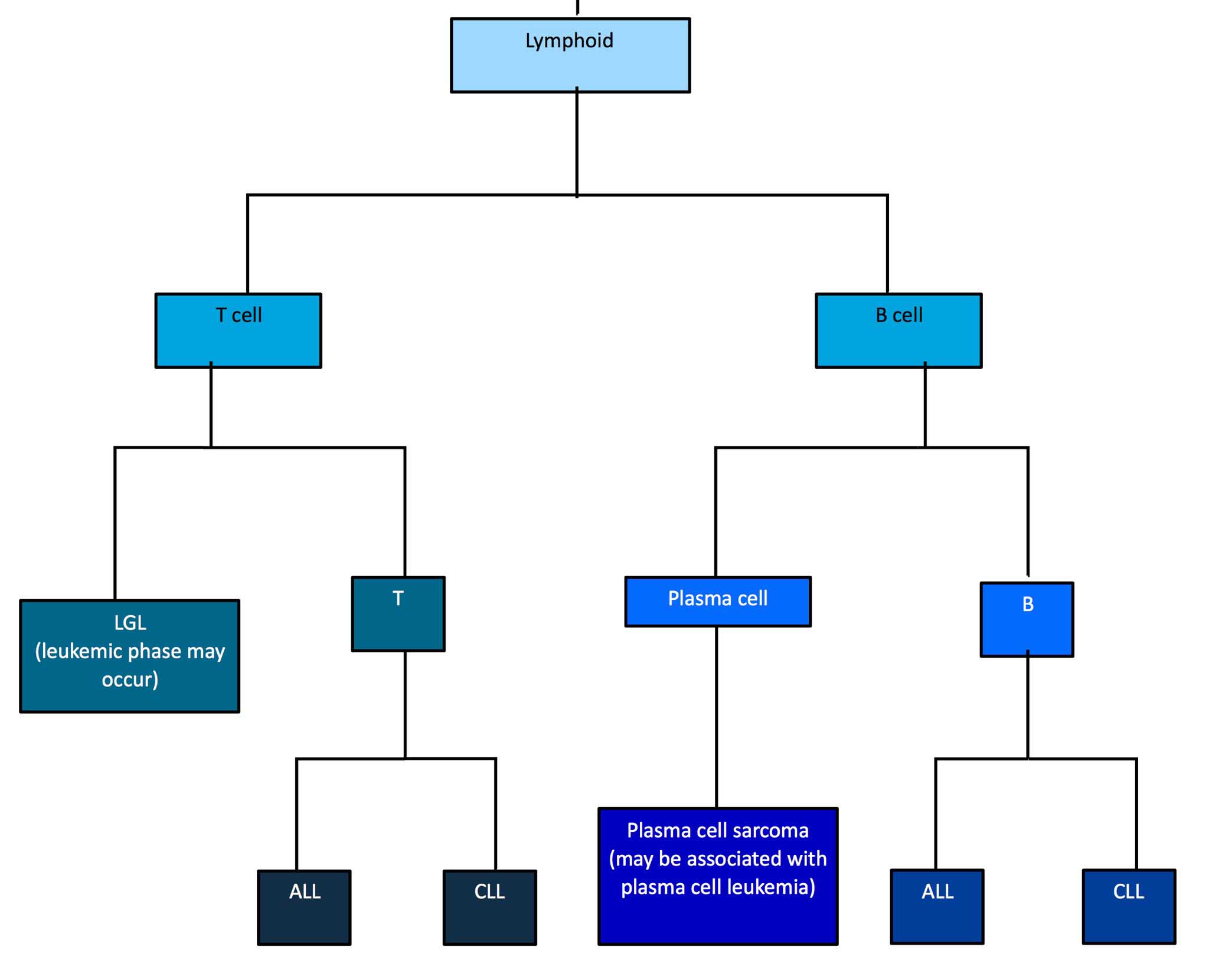
Hemopoietic neoplasms include clonal proliferations of both lymphoid and myeloid cells (Fig. 3.1). Lymphoid neoplasms occur much more frequently than myeloid neoplasms in veterinary medicine. Myeloid refers to bone marrow-derived hemopoietic cells, with the exception of lymphocytes, as these may originate from tissues such as spleen, liver, thymus, and lymph nodes in addition to bone marrow. Accurate identification and classification of hemopoietic neoplasms aid the clinician in prognostication and in recommending appropriate treatment options for affected patients. Classification schemes for canine and feline hemopoietic neoplasms have generally been modified from human medicine. Hemopoietic neoplasms in other domestic animals have received less attention. Phenotyping of human hemopoietic neoplasms relies, in part, on sophisticated molecular and genetic tests which are not always available in veterinary medicine. However, great advances have been made in diagnosing, treating, and understanding the pathophysiology of these neoplasms in veterinary medicine, much to the credit of studies carried out by our counterparts in human medicine.


Leukemia may be present with both lymphoid and myeloid neoplasms and generally refers to the presence of bone marrow-derived neoplastic cells in the peripheral blood. In other instances, where the primary hemopoietic neoplasm is a solid tumor (e.g. of lymphocytes, plasma cells, or mast cells) that develops extramedullary (outside the bone marrow) but releases neoplastic cells into the circulation, the term “leukemic phase” of the neoplasm is used. Confusion may occur with these definitions since, at the time of diagnosis, it is not always possible to determine whether the bone marrow or the mass represents the primary or metastatic site.
The presence of leukemia is relatively easy to determine when neoplastic cells occur in high numbers in the circulation, cell morphology is abnormal, and there is no physiologic explanation for the peripheral blood findings. Diagnosis can be more difficult when only low numbers of morphologically abnormal neoplastic cells are present in the peripheral blood. In other cases, the neoplastic cells may be proliferating in the bone marrow without being released into the peripheral blood, a situation that may be called preleukemia. Cytopenia of one or more non-neoplastic hemopoietic cell lines may accompany hemopoietic neoplasia and bone marrow examination is indicated, particularly if the cytopenia is unexplained because neoplastic cells are not identified in the peripheral blood.
Referring to lymphocytes and tissues where lymphocytes develop.
Can have different hematologic meanings: 1. referring to all hemopoietic cells in the bone marrow except lymphocytes, 2. referring to all bone marrow leukocytes except lymphocytes.
Mononuclear, non-phagocytic leukocyte responsible for humoral (B lymphocyte) and cell-mediated (T lymphocyte) immune responses.
Organ responsible for production of hemopoietic cells; found in the medullary cavity, especially the ends of long bones (e.g. femur) and flat bones (e.g. the pelvis, sternum.)
Presence of bone marrow-derived neoplastic cells in peripheral blood.
Terminally differentiated B lymphocyte that secretes specific antibody.
Mononuclear, granular leukocyte important in hypersensitivity reactions.
Term describing the absence of neoplastic hemopoietic cells in peripheral blood despite the presence of neoplastic hemopoietic cells in bone marrow.
Abnormal uncontrolled growth of cells that are unresponsive to normal physiologic growth controls; may be benign or malignant.
General term for a decrease in number of any cell line in peripheral blood.

