See Fig. 7.3 – 7.18 for various urine sediment findings.
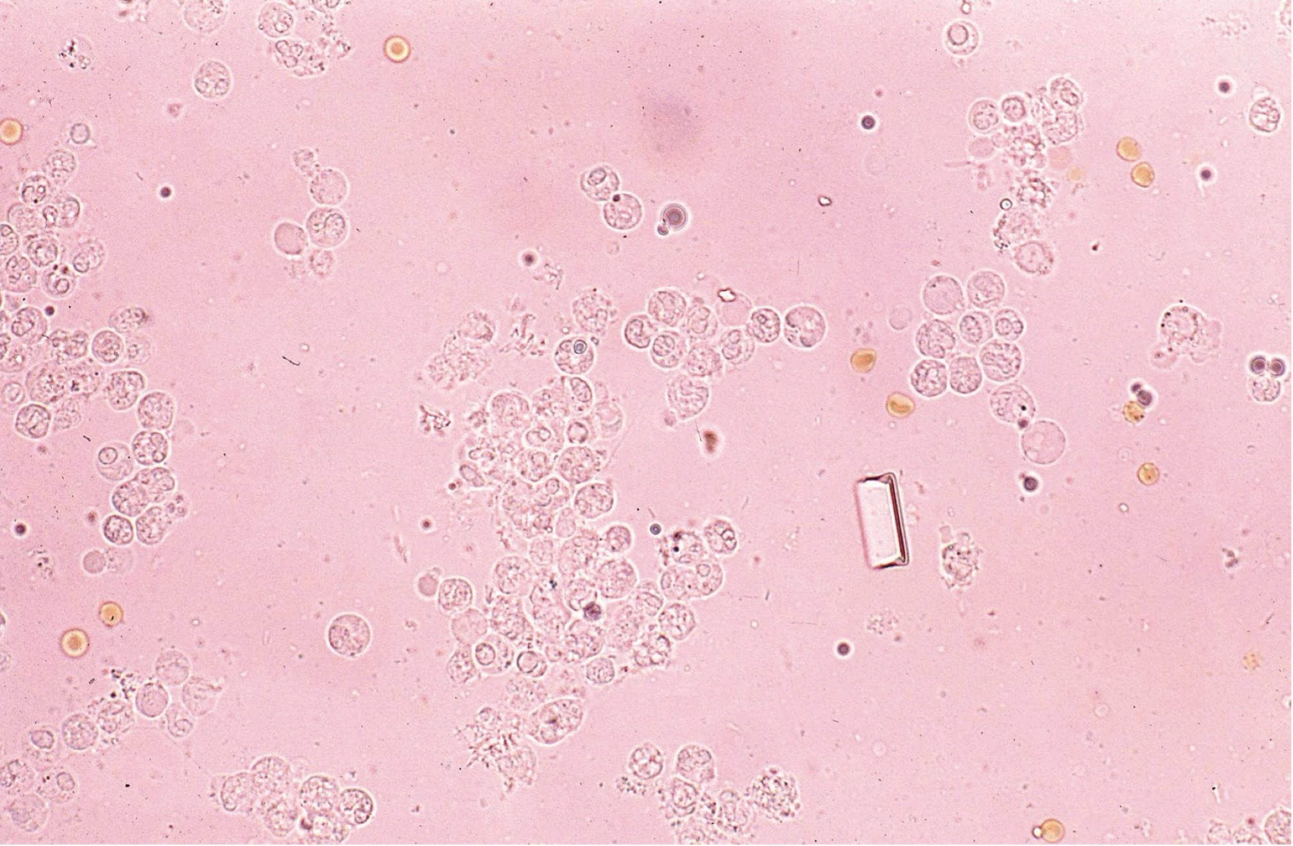 Figure 7.3 Urine sediment, unstained. Numerous leukocytes and erythrocytes; single struvite crystal.
 Figure 7.4 Urine sediment, sedistain. Numerous erythrocytes and leukocytes.
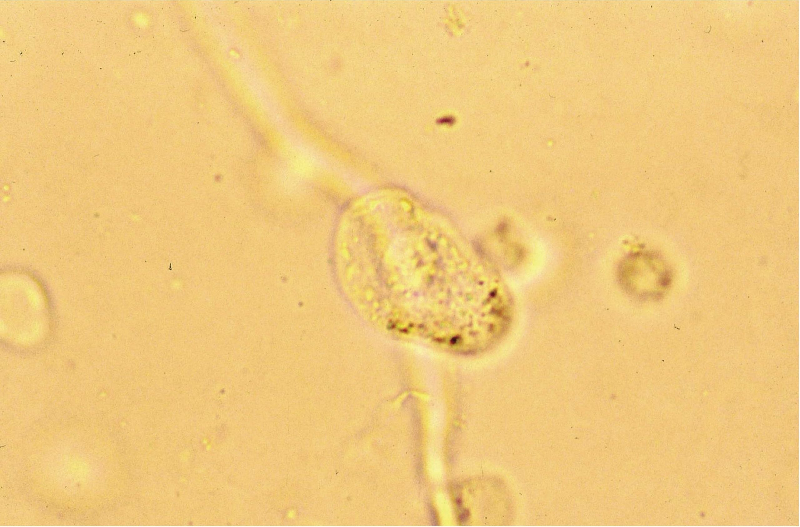 Figure 7.5 Urine sediment, unstained. Squamous epithelial cell.
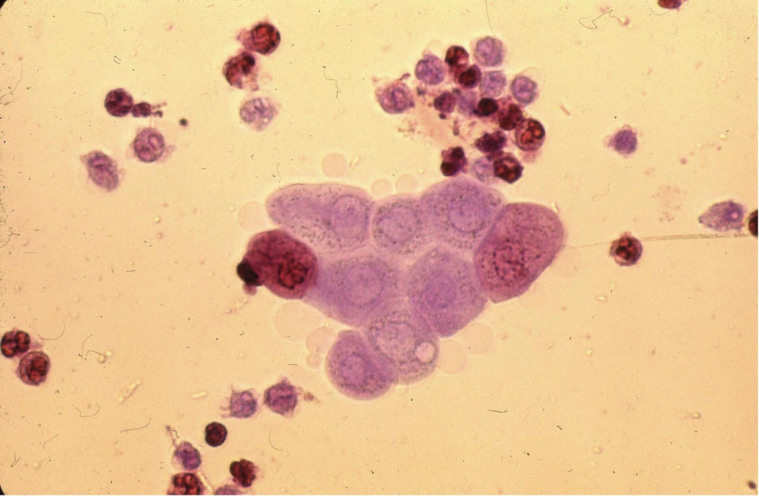 Figure 7.6 Urine sediment, sedistain. Cluster of transitional epithelial cells with leukocytes.
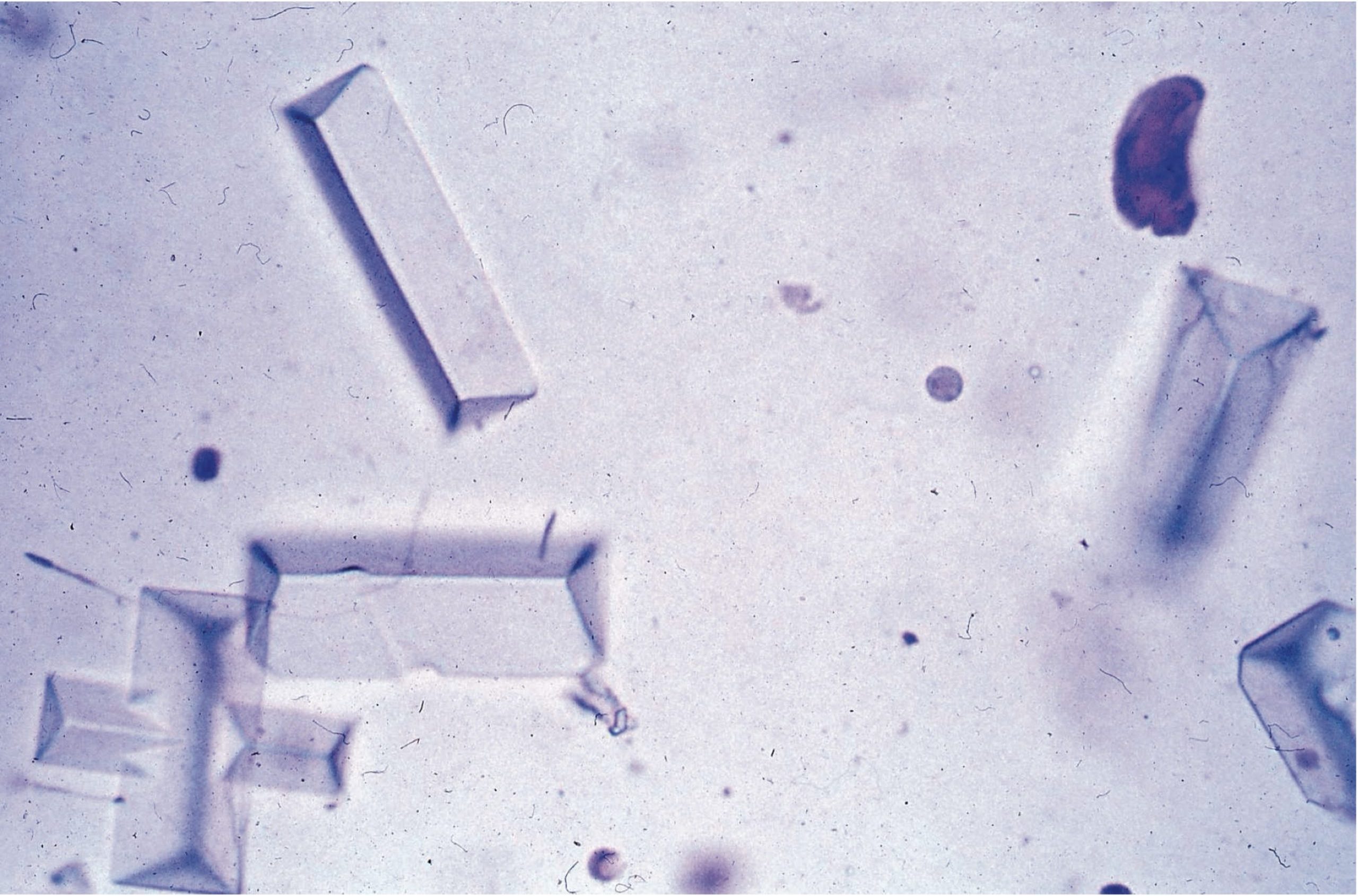 Figure 7.7 Urine sediment, sedistain. Struvite crystals.
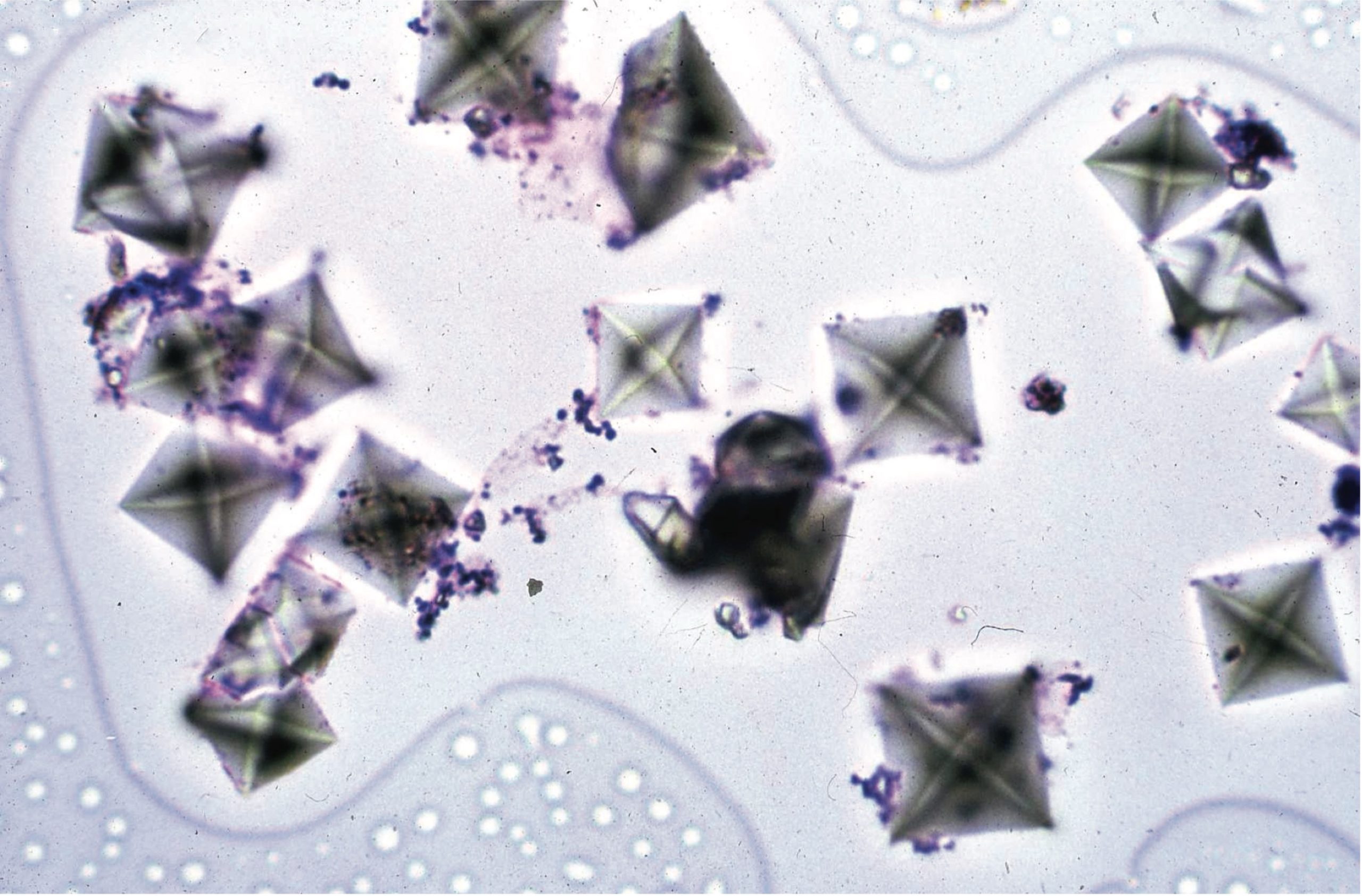 Figure 7.8 Urine sediment, sedistain. Calcium oxalate dihydrate crystals.
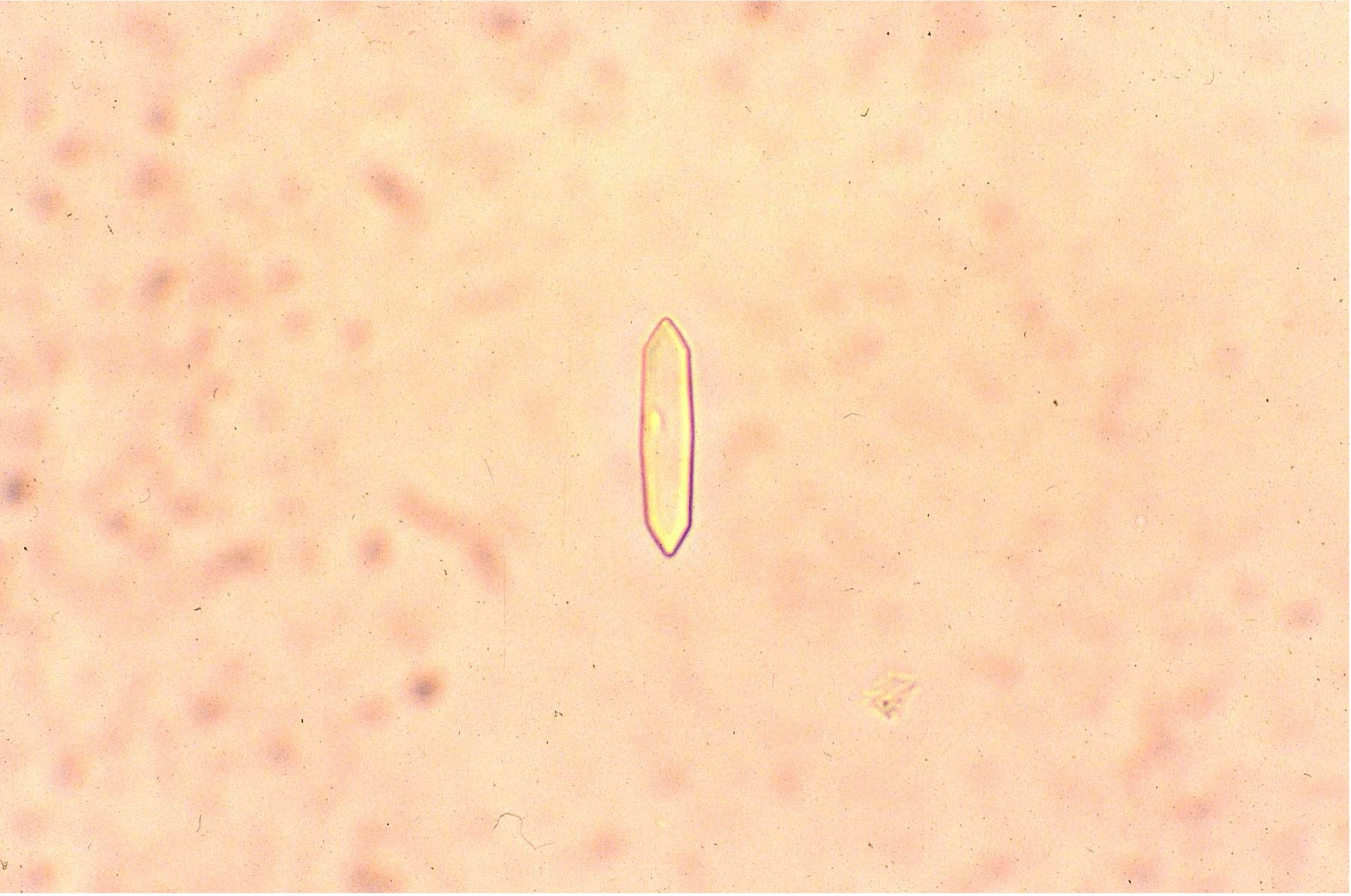 Figure 7.9 Urine sediment, unstained. Calcium oxalate monohydrate crystals.
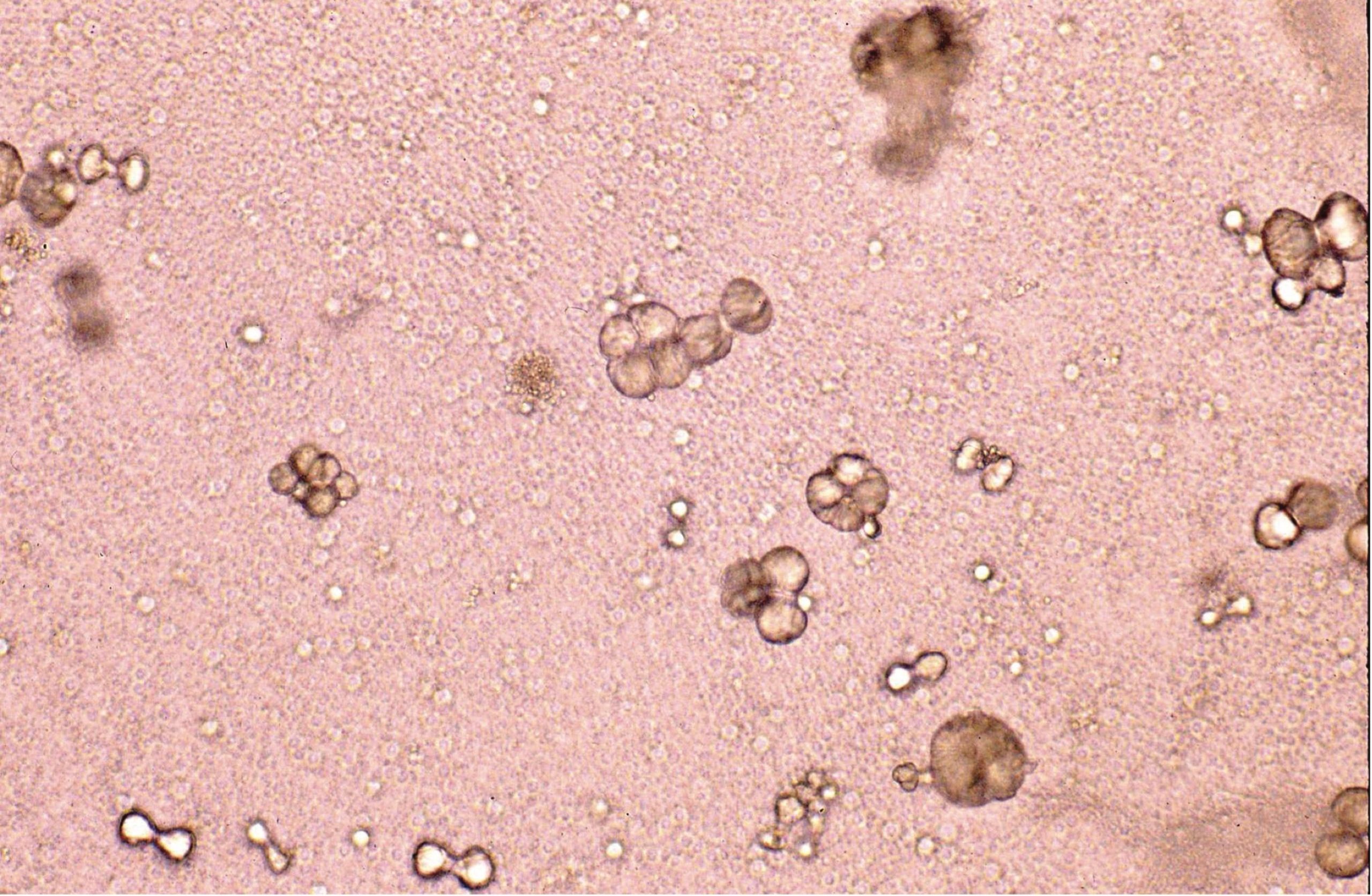 Figure 7.10 Urine sediment, unstained. Calcium carbonate crystals. May be spherical to dumbbell-shaped.
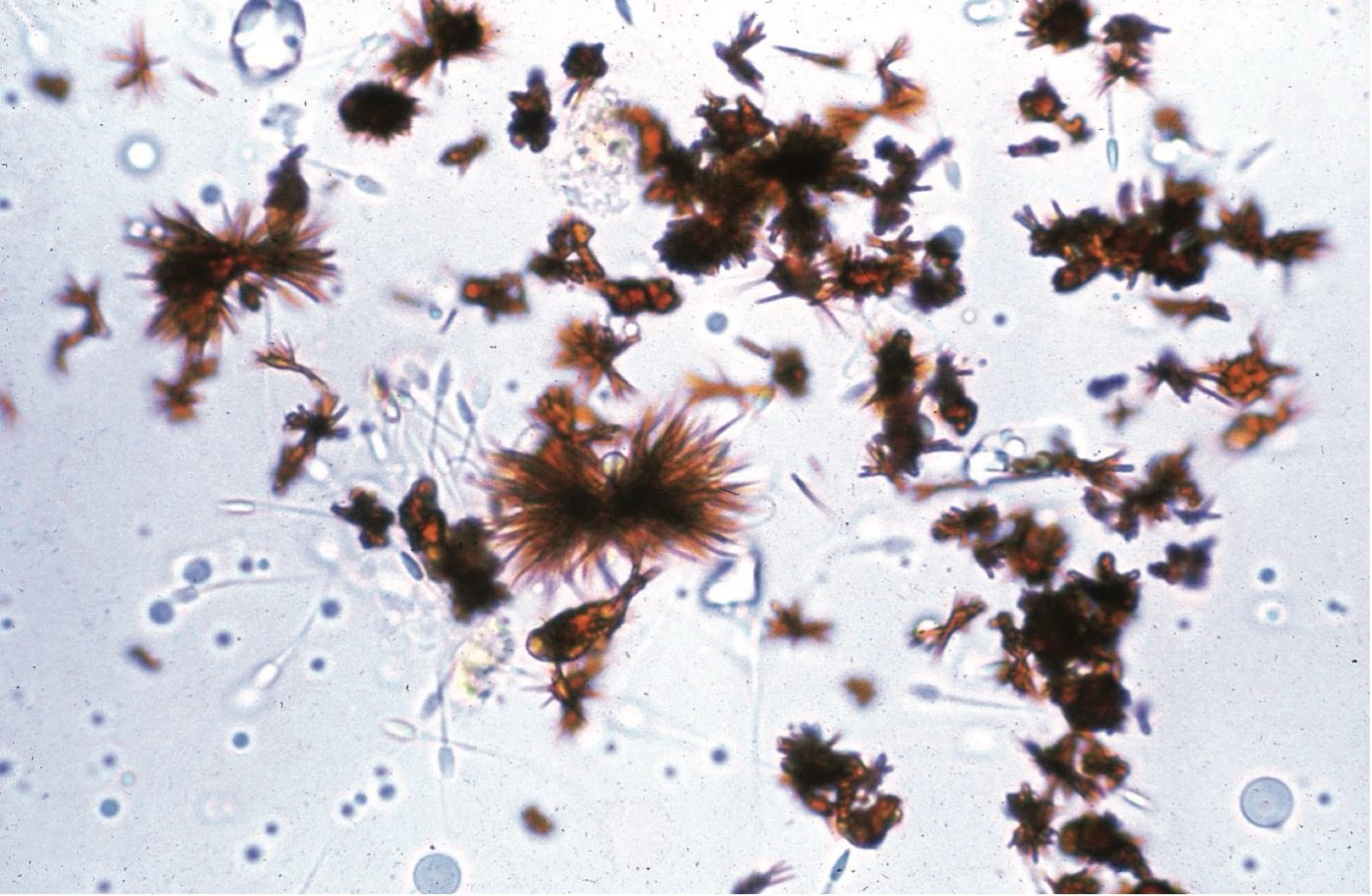 Figure 7.11 Urine sediment, unstained. Bilirubin crystals. These are yellow-brown. Note also spermatozoa.
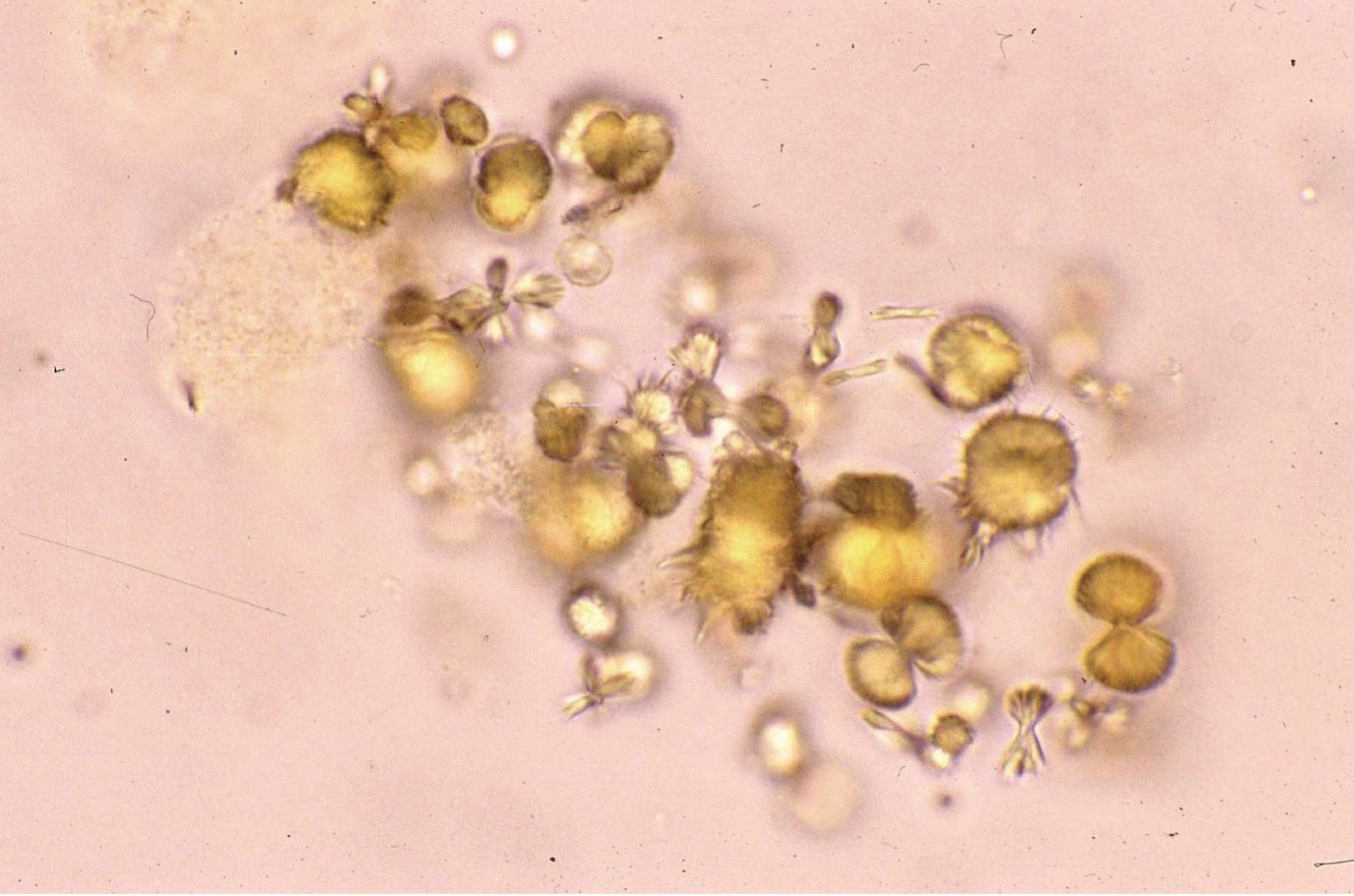 Figure 7.12 Urine sediment, unstained. Ammonium biurate (round with spikes) and urate crystals.
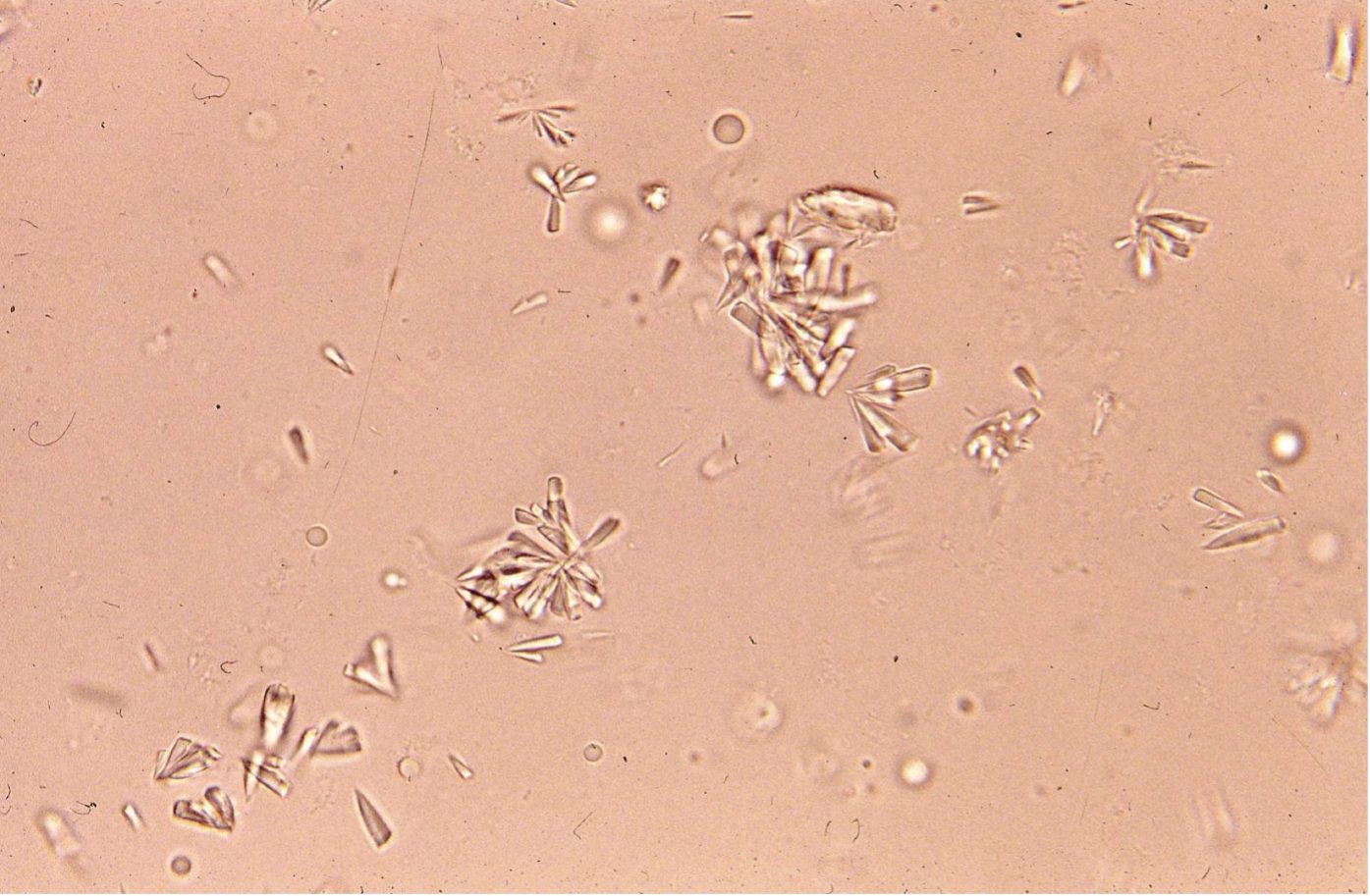 Figure 7.13 Urine sediment, unstained. Calcium phosphate crystals.
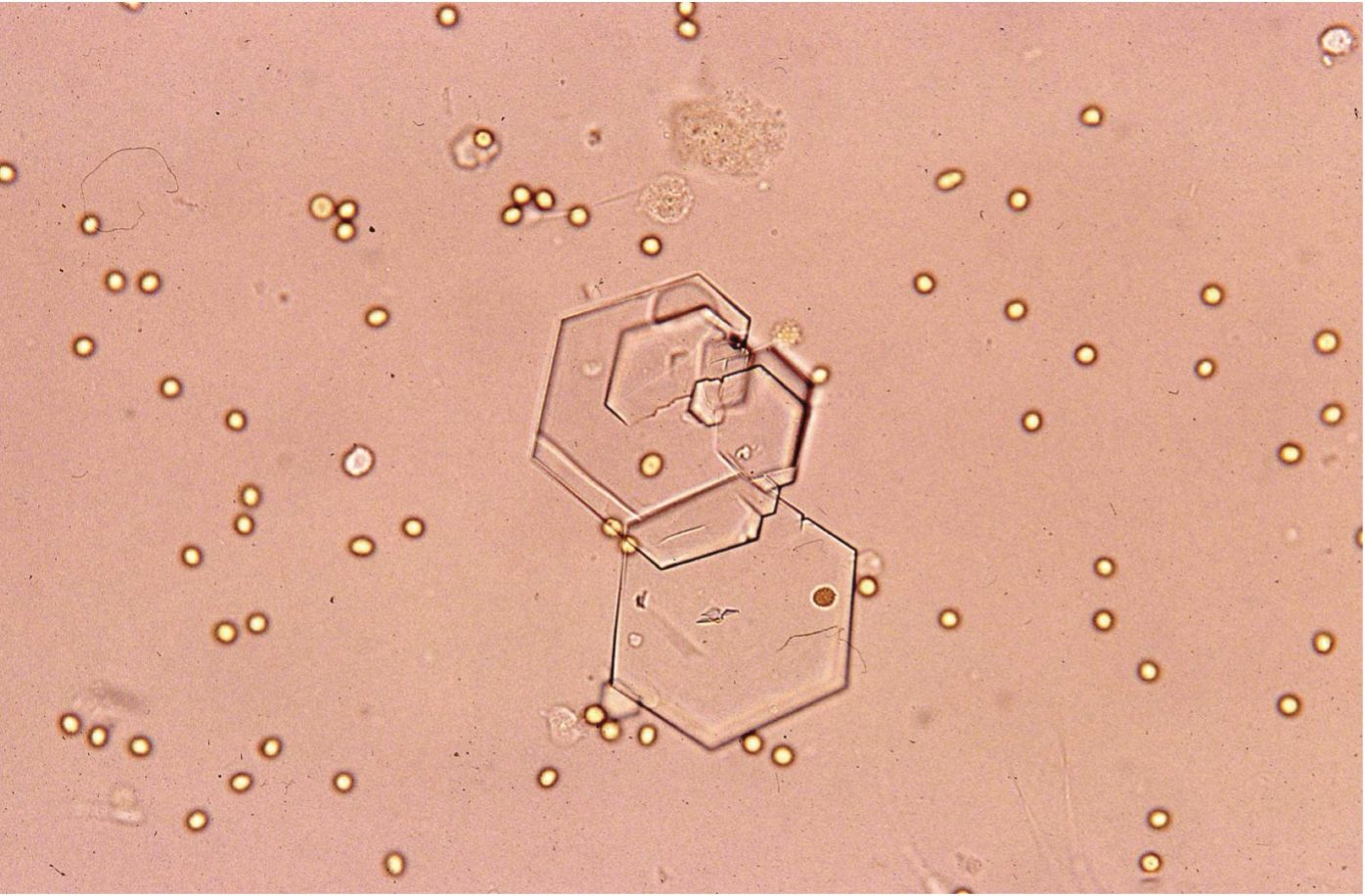 Figure 7.14 Urine sediment, unstained. Cystine crystals.
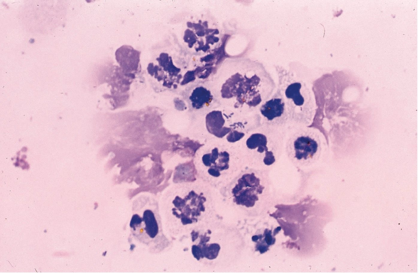 Figure 7.15 Urine sediment, Wright-Giemsa. Degenerate neutrophils. Intracellular rods are present in the central neutrophil.
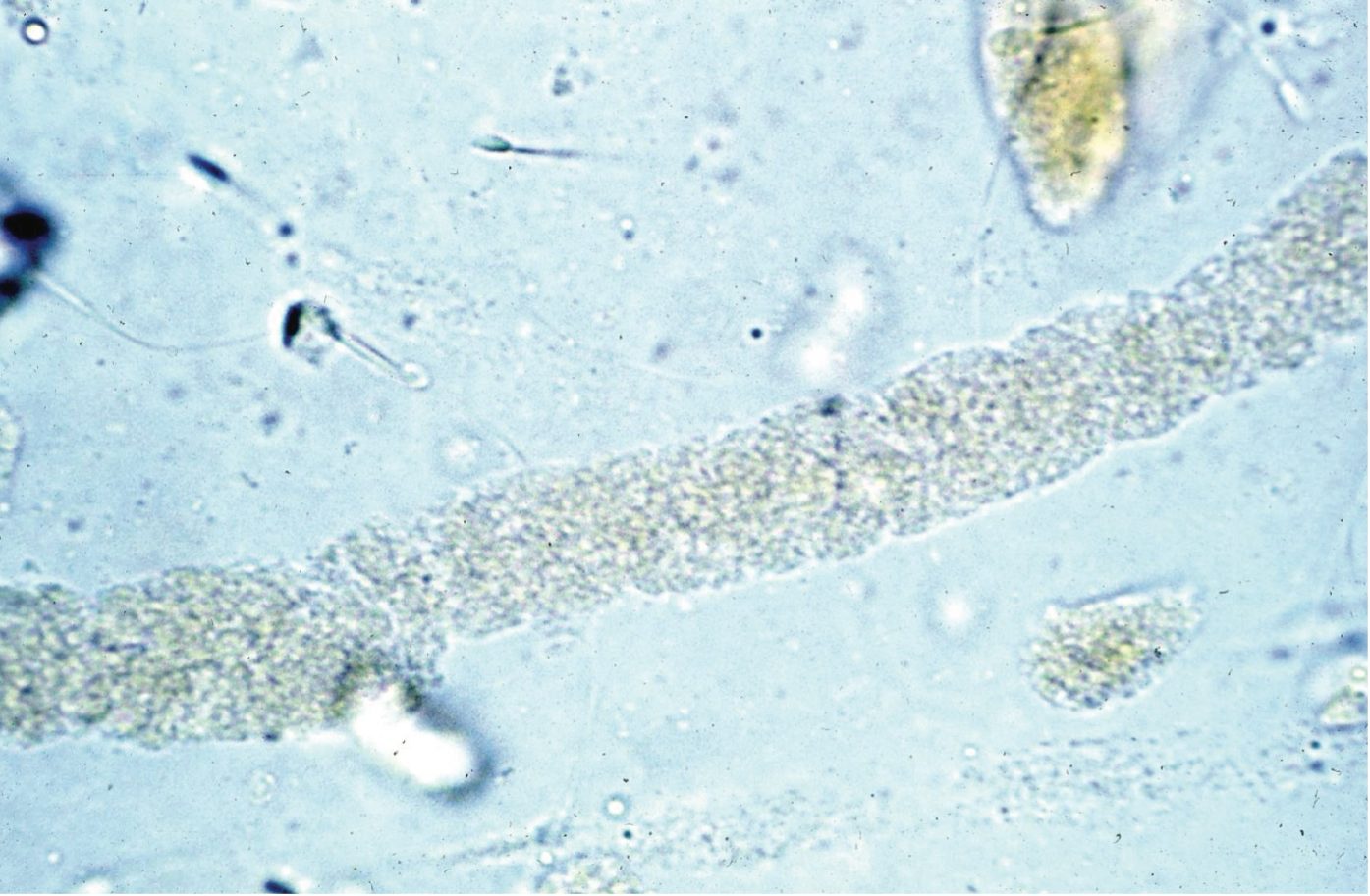 Figure 7.16 Urine sediment, unstained. Granular cast.
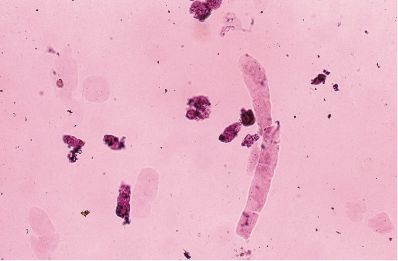 Figure 7.17 Urine sediment, sedistain. Hyaline cast.
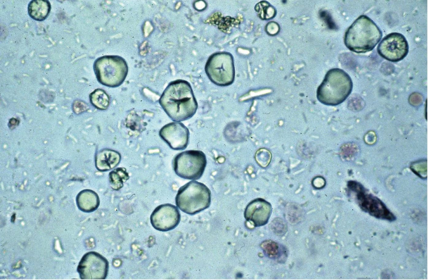 Figure 7.18 Urine sediment, sedistain. Starch granules (artifact), from glove powder.

