Transudates
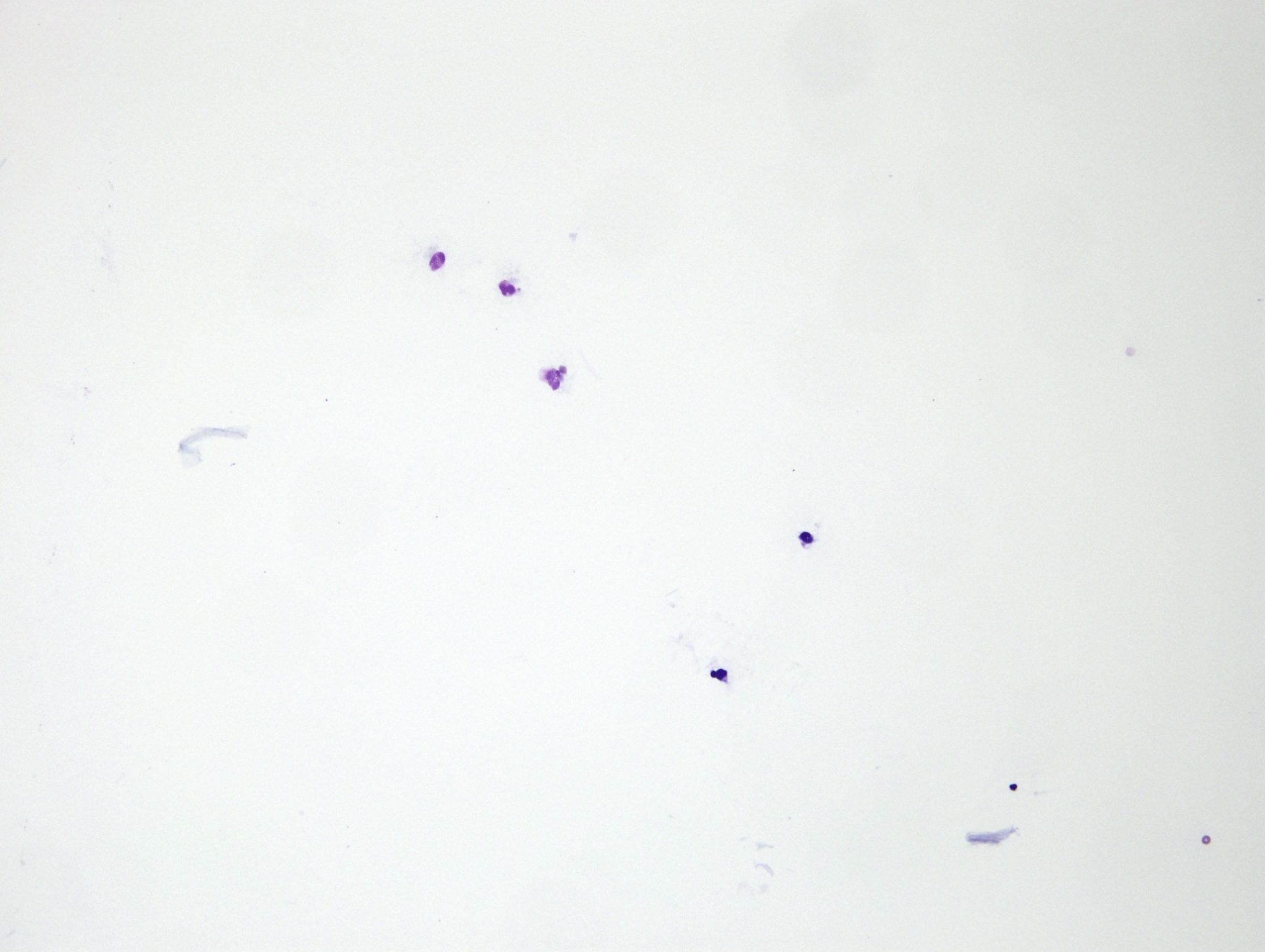
A transudate is a very low protein, cell poor fluid (protein <25 g/L; nucleated cell count <1.5 x 10⁹/L). Transudates are clear and colorless and cells are difficult to locate on direct smears (Fig. 5.31; see also Video Cytology fluid handling). Many laboratories use a cytospin centrifuge to prepare concentrated cytological preparations of fluid samples, particularly those that are cell poor. This instrument preserves cell morphology and concentrates all the cells from a certain volume of fluid (up to 500 µL) into a circular dot (about 5 mm in diameter) on the slide.

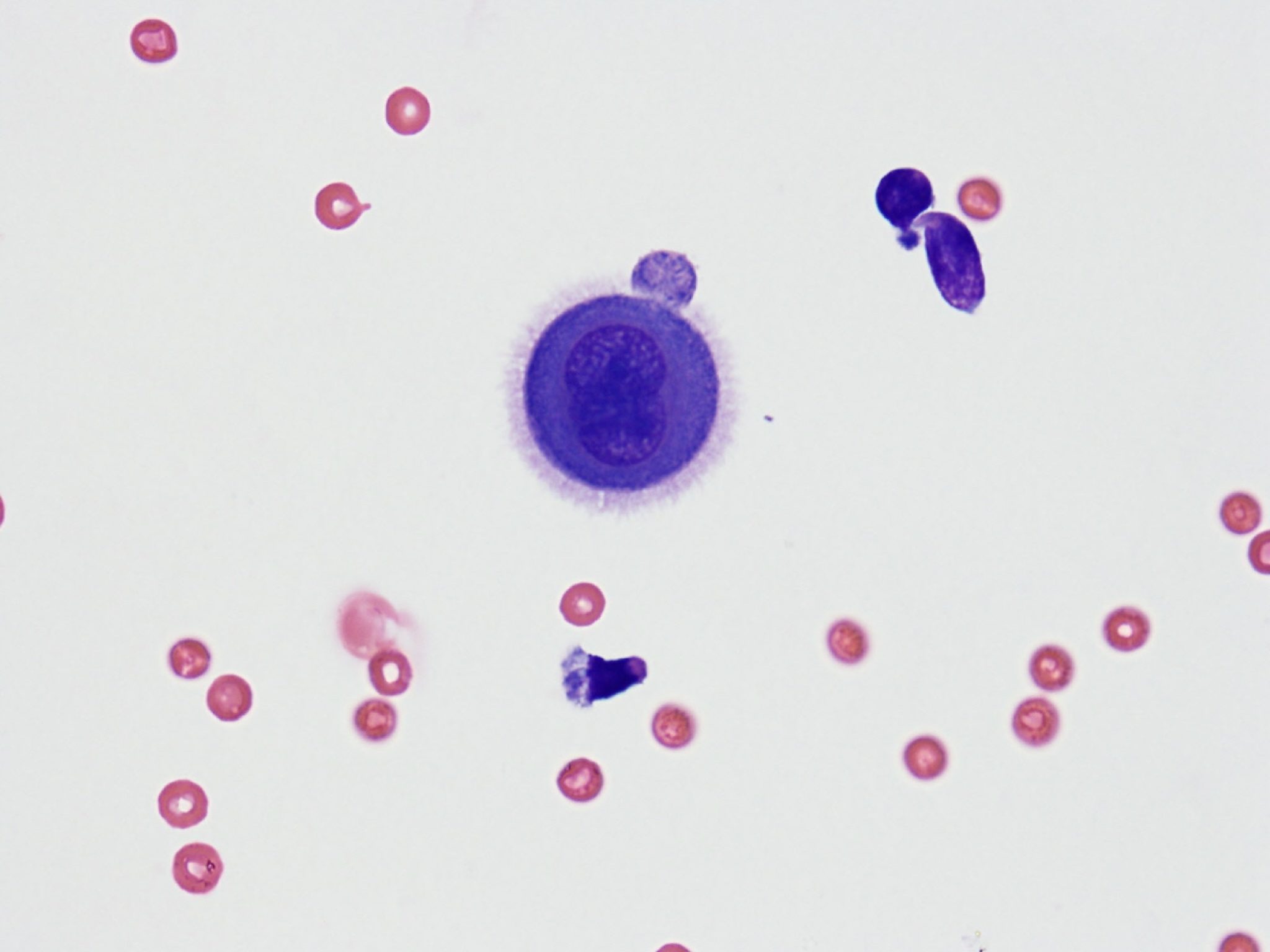
Transudates form as a result of decreased plasma oncotic pressure (due to marked hypoalbuminemia <10 g/L), increased capillary hydrostatic pressure, lymphatic obstruction, or any combination of the three. Nucleated cells are often a mix of macrophages, nondegenerate neutrophils, and mesothelial cells. One should not assume that inflammation is present simply because neutrophils are present. The total nucleated cell count is important in interpreting the significance of neutrophils in a sample because neutrophils form a proportion of the cell population in many normal fluids. For example, normal equine peritoneal fluid contains 0.5-9.0 x 10⁹/L (usually <4.0 x 10⁹/L) nucleated cells, consisting of about 50% neutrophils and 50% macrophages. Mesothelial cells line the pleural, peritoneal, and pericardial surfaces (Fig. 5.32). With excess fluid accumulation in body cavities, mesothelial cells become activated, slough from the lining surface, proliferate by mitosis, and have the ability to phagocytose cellular and particulate material.

Certain neoplasms may interfere with venous and lymphatic drainage, resulting in transudate development. However, if neoplastic cells are actually exfoliating into the body cavity, the fluid is usually a modified transudate. Chronic passive congestion associated with heart failure can, at least initially, result in transudate formation. Later, the fluid may become blood-tinged and protein concentration and cell counts will increase, producing a modified transudate.
Very low protein, cell poor fluid.
Cell lining body cavities that may exfoliate into body cavity fluids when there is inflammation or accumulation of excess fluid for other reasons; mesothelial cells can become phagocytic.
Transudate that has been modified by the presence of additional protein, cells, or both.

