"

Spindle Cell Tumors

Mesenchymal spindle cell tumors may not exfoliate well, as cells are embedded in extracellular matrix. Cells are generally individual rather than adherent, fusiform, and have indistinct cell borders. Nuclei are often elongate and cytoplasmic tails may fade into the background. Differentiation according to tissue of origin is sometimes possible- for example, there may be evidence of collagen, cartilage, bone, fat, or myxomatous material formation by the tumor cells. In general, benign spindle cell tumors are indicated by the suffix “-oma”, for example, fibroma; malignant mesenchymal tumors are called sarcomas, for example, fibrosarcoma (see Fig. 5.14 for well differentiated spindle cells and Fig. 5.15 for spindle cells with malignant features).
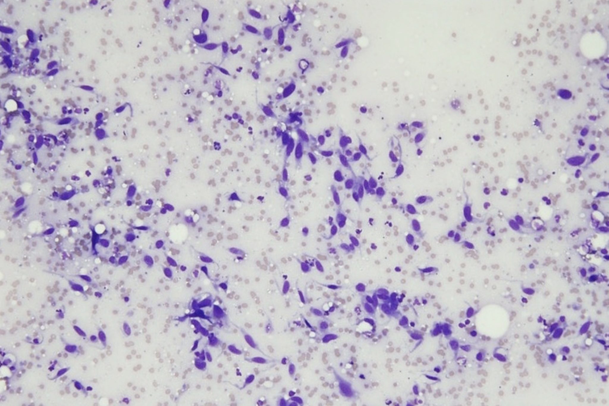
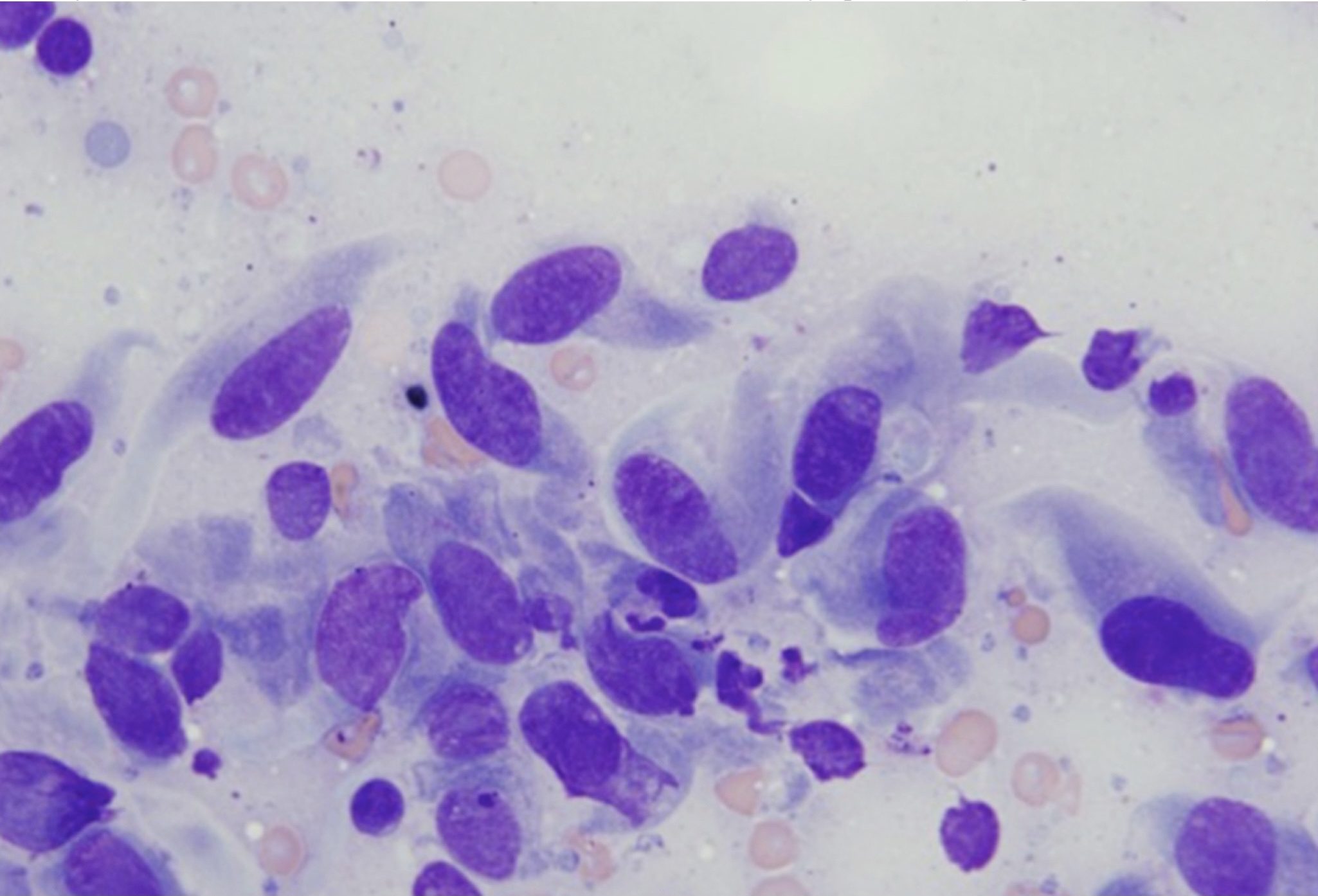
Figure 5.14 Well differentiated spindle cells. The cells are uniform in size and shape. (Wright-Giemsa; LP.) (Image courtesy of Dr. Angelica Galezowski.)
Figure 5.15 Spindle cells with malignant features. The cells are large and often binucleate. These are leiomyosarcoma cells that have metastasized to the mandibular lymph node. (Wright-Giemsa; HP oil.)
definition

License

Icon for the Creative Commons Attribution-NonCommercial-NoDerivatives 4.0 International License

Veterinary Clinical Pathology: An Introduction Copyright © by Marion Jackson; Beverly Kidney; and Nicole Fernandez is licensed under a Creative Commons Attribution-NonCommercial-NoDerivatives 4.0 International License, except where otherwise noted.