Round Cell Tumors
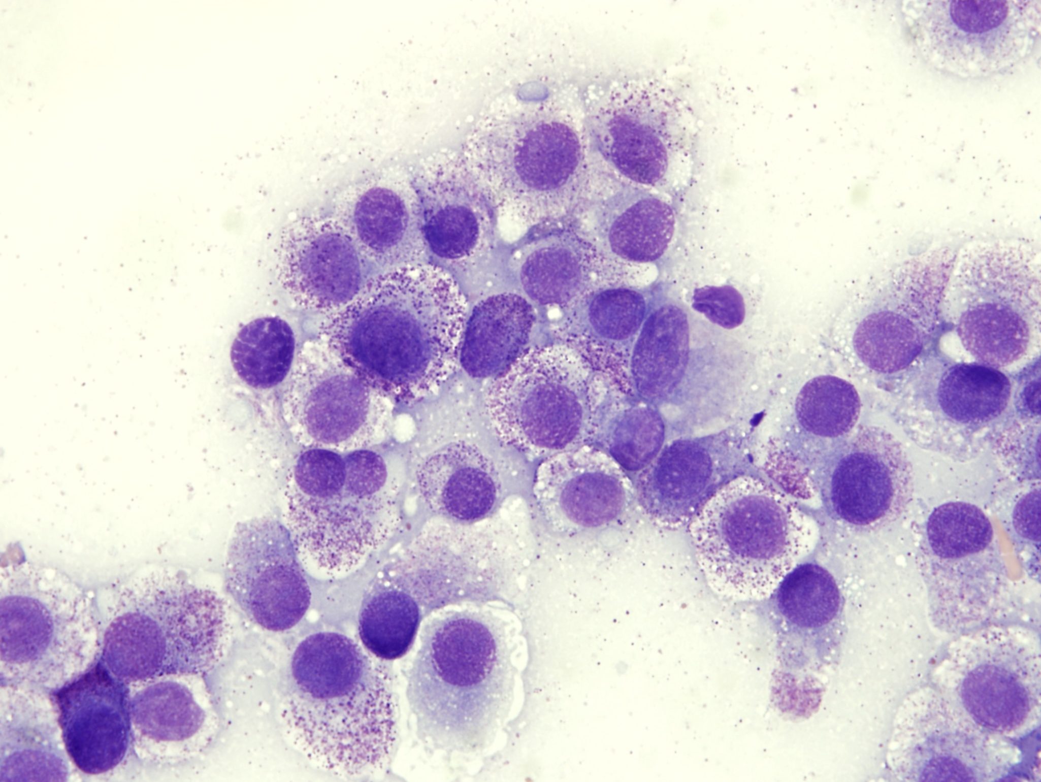
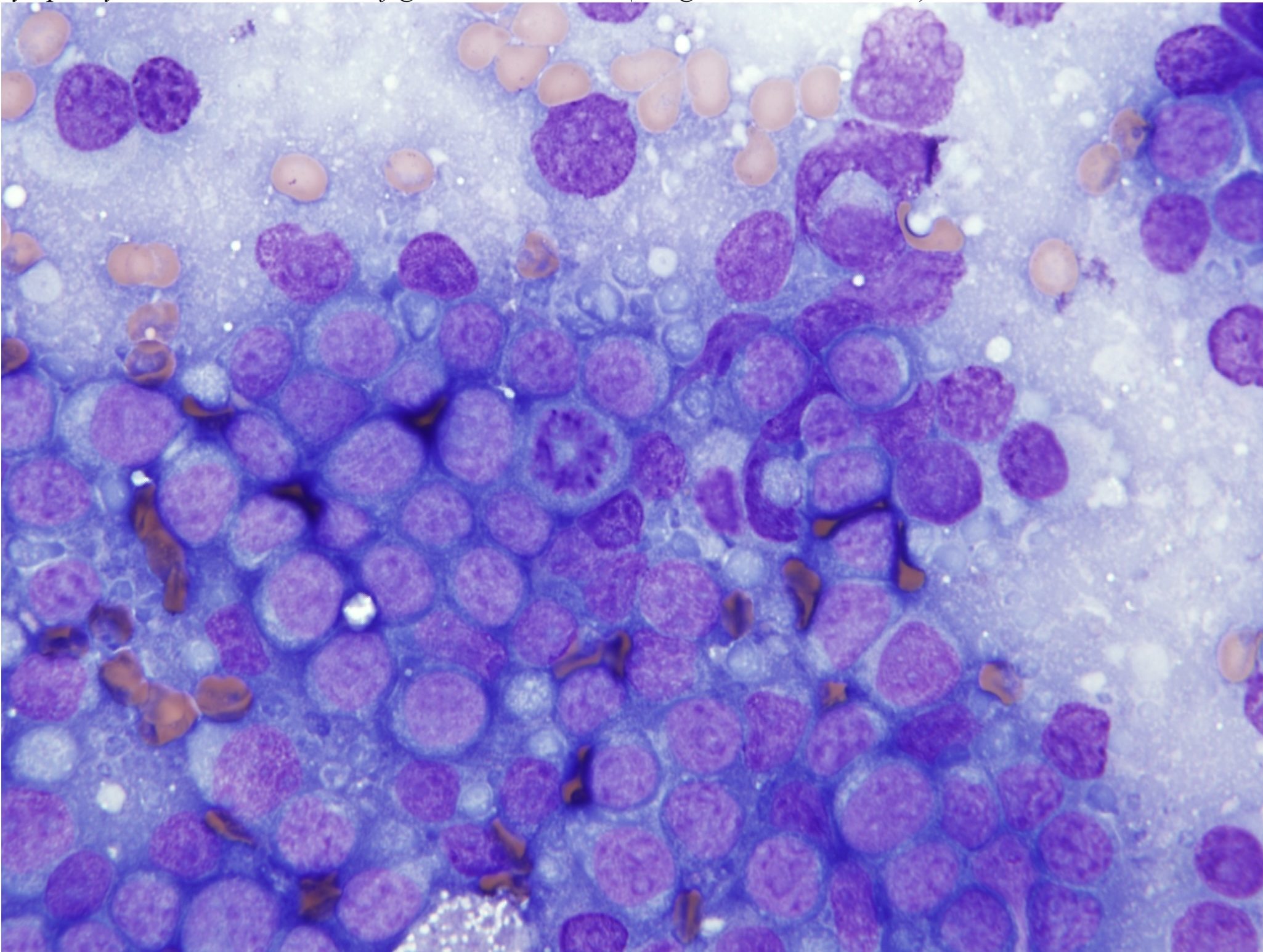
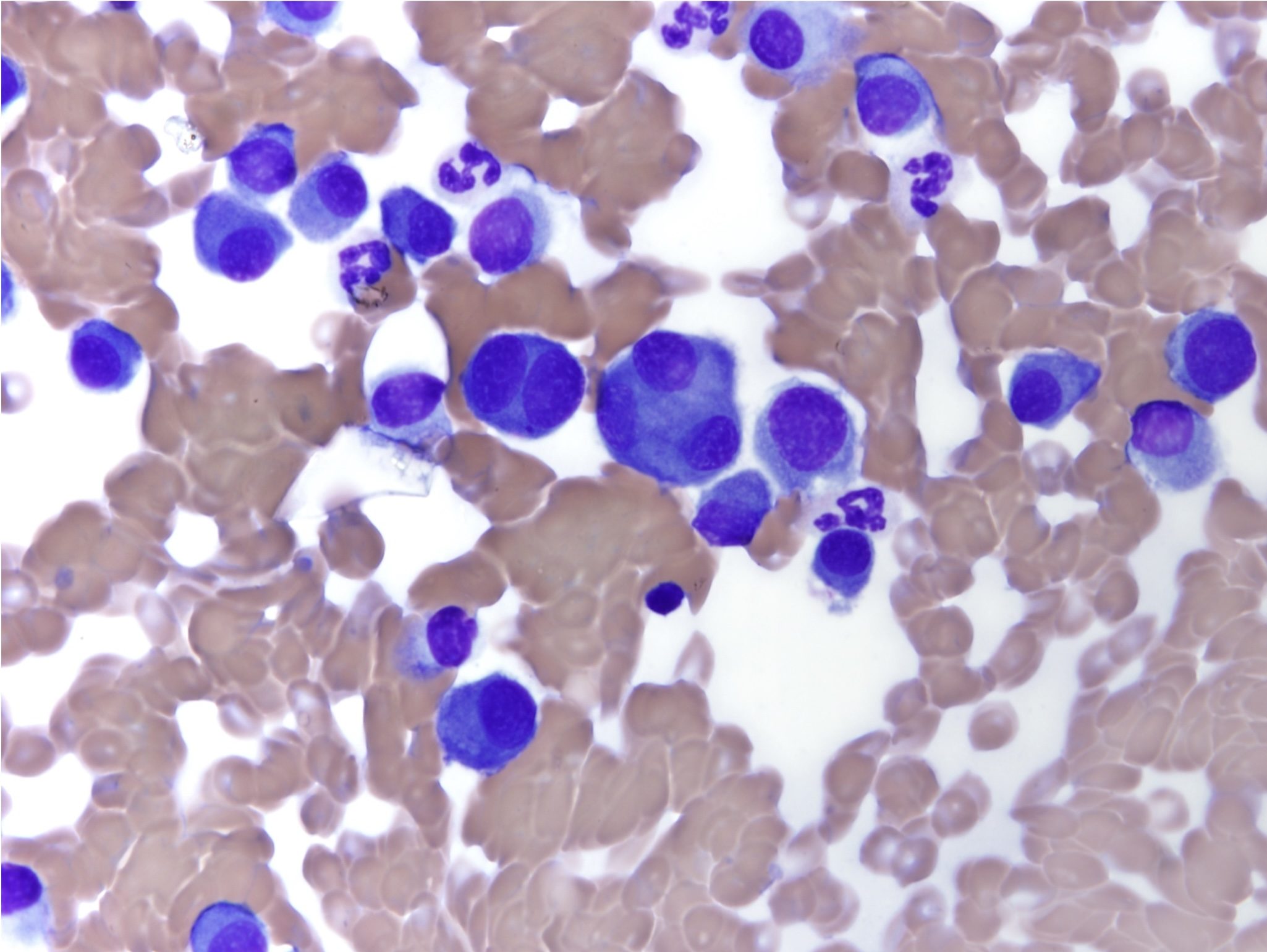
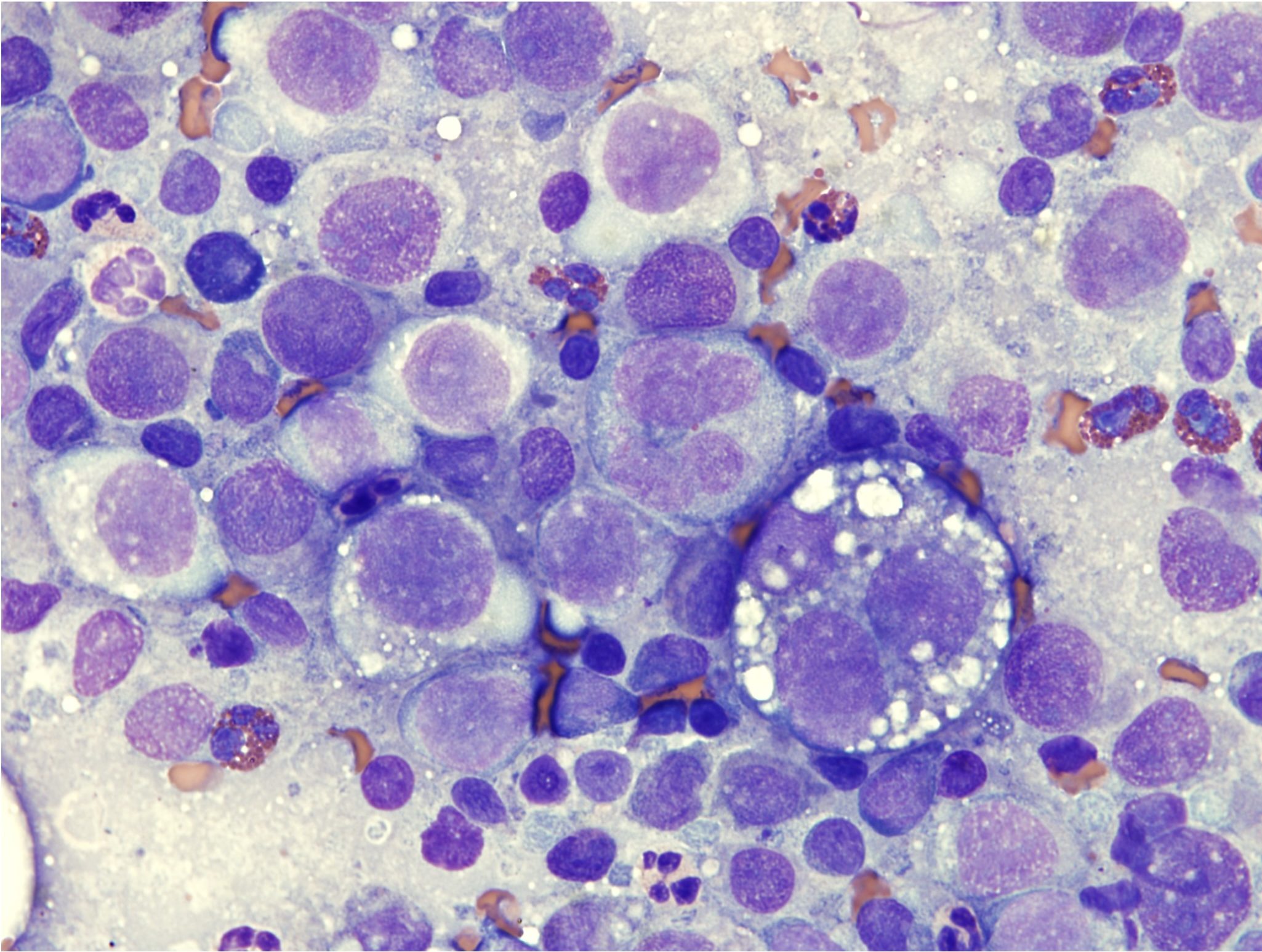
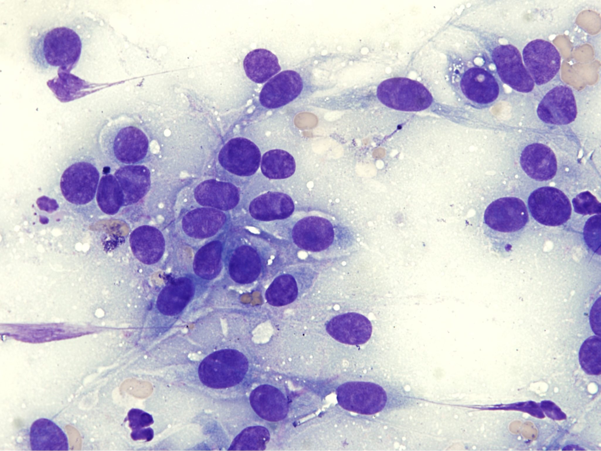
Although mesenchymal discrete round cells include all hemopoietic cell lines, those cells that form solid masses include only mast cells, lymphocytes, plasma cells and histiocytes. Transmissible venereal tumor, a canine round cell tumor of unknown tissue origin, is included in the discrete round cell group due to its morphology. Round cell tumors generally exfoliate well. Cells have distinct cell borders and are individual rather than cohesive. Often there is sufficient differentiation to diagnose these based on cytology alone, although additional tests are sometimes needed. Mast cells are generally easy to recognize on cytology as the cytoplasm contains numerous purple granules (Fig. 5.18). Lymphocytes resemble lymphocytes from peripheral blood, but may be large and have increased amounts of cytoplasm (Fig. 5.19). Plasma cells typically have an eccentric nucleus with a clear Golgi zone and deeply basophilic cytoplasm (Fig. 5.7). Histiocytes have pale cytoplasm with round to indented nuclei, and lack any of the features described in other round cells (Fig. 5.20).
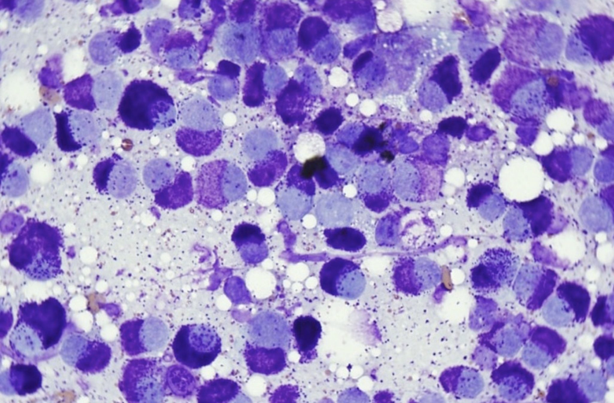
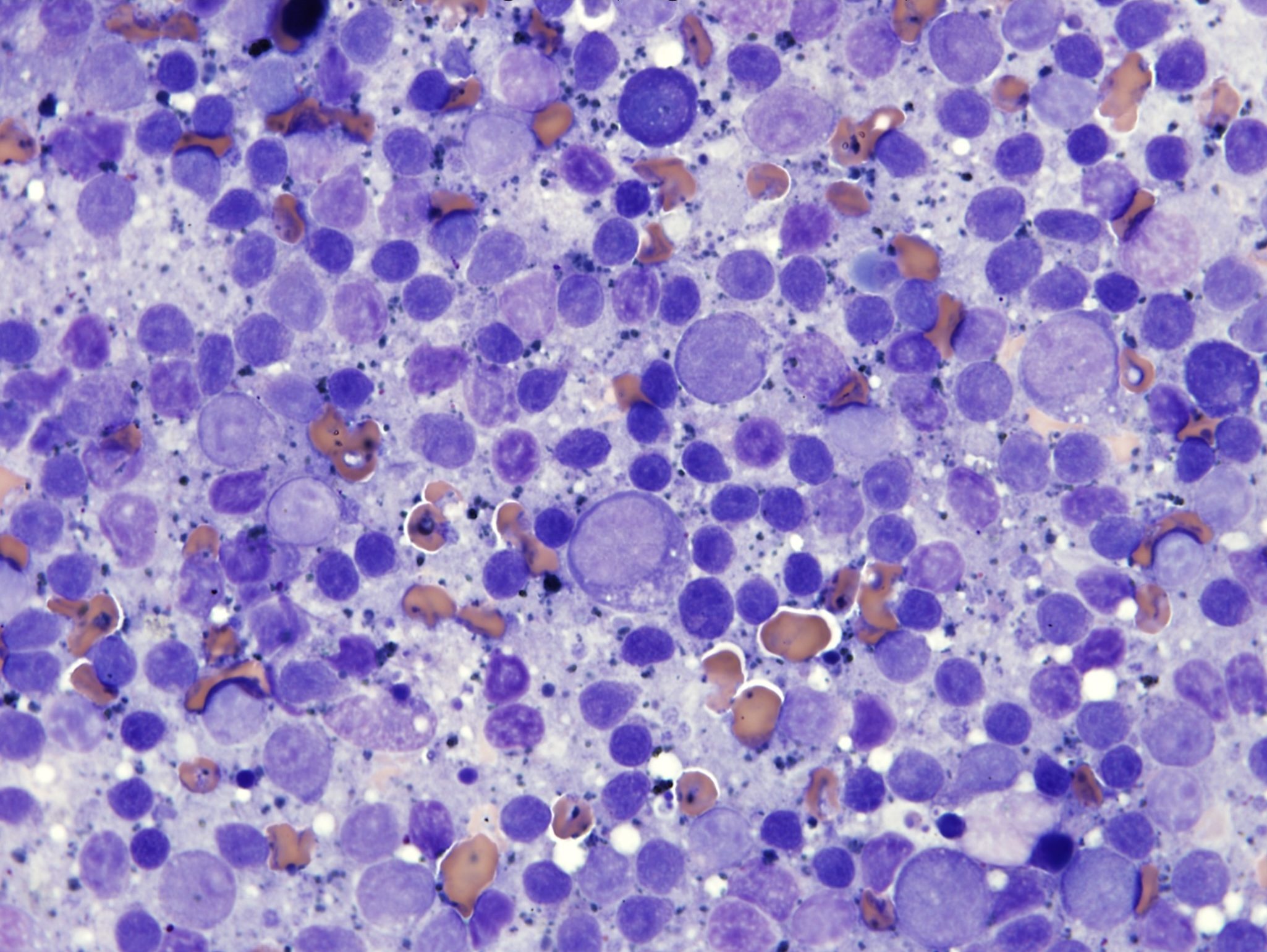
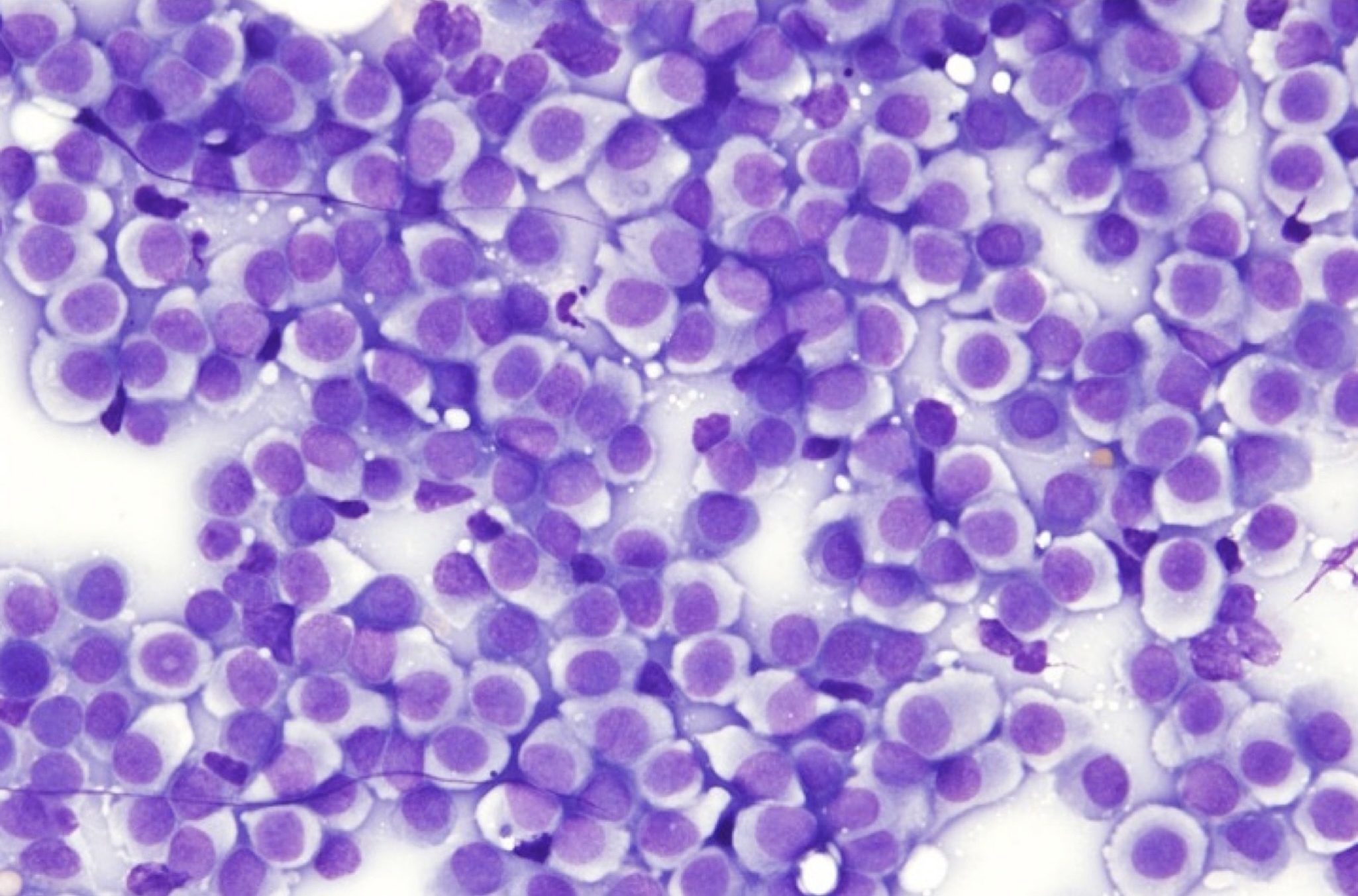
Distinguishing between benign and malignant tumors within this group cannot always be done based on morphology. They do not display the typical criteria of malignancy of carcinomas and sarcomas as described above, with the possible exception of histiocytic sarcoma. Pattern of growth, local lymph node involvement, evidence of metastasis, and published studies assist the pathologist and clinician in assessing the health risk of these tumors in individual animals. For example, poorly granulated mast cell tumors (Fig. 5. 21) are more likely to behave in a malignant manner than those that are well differentiated and heavily granulated (see Fig. 5.6 and Fig. 5.18 for well differentiated mast cells). However, mast cell tumors are all potentially malignant, regardless of morphology, and histologic grading is recommended to further evaluate the prognosis. Lymphoid tumors are always malignant, therefore are correctly called lymphosarcoma (Fig. 5.22), but must be distinguished from accumulations of reactive lymphocytes, for example, due to vaccine reactions. Plasma cell tumors can be solitary and carry a good prognosis with surgical removal (Fig. 5.23), or they can be part of a systemic malignant plasma cell tumor. Histiocytomas (cutaneous tumors of histiocytes; Fig. 5.20) are benign and may spontaneously regress in young dogs despite occasional pleomorphism and high numbers of mitoses, whereas histiocytic sarcomas display many criteria of malignancy including marked anisocytosis, anisokaryosis, and multinucleation; frequently occur in internal organs; and have a high metastatic potential (Fig. 5.24).
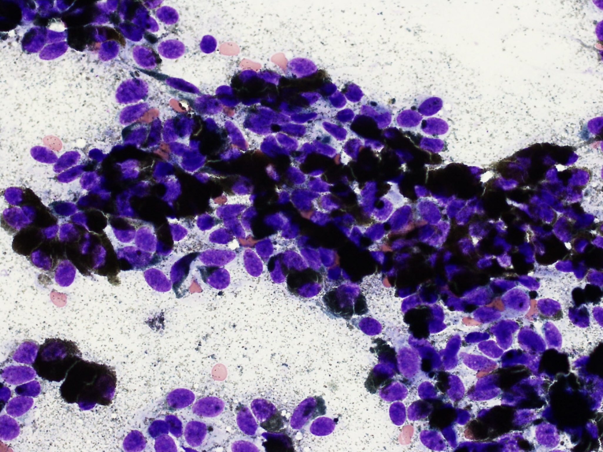
Melanomas and malignant melanomas are not easily categorized. Melanocytes are derived from the neuroectoderm and, as such, should be considered as epithelial in origin. The diagnosis is less challenging when melanin granules are present (Fig. 5.25). However, pigmented cells may be present in very low numbers, and malignant melanocytes do not always form visible melanin granules (Fig. 5.26). In addition, malignant melanocytes can resemble epithelial, spindle, or round cells, alone or in combination. If the pathologist is having great difficulty in deciding the origin of a tumor, amelanotic malignant melanoma should be considered.
Tissue macrophage.
Referring to lymphocytes and tissues where lymphocytes develop.
Solid malignant lymphoid tumor.
Immunologically stimulated lymphocyte that typically has darkly staining cytoplasm

