Pituitary Gland and Adrenal Glands
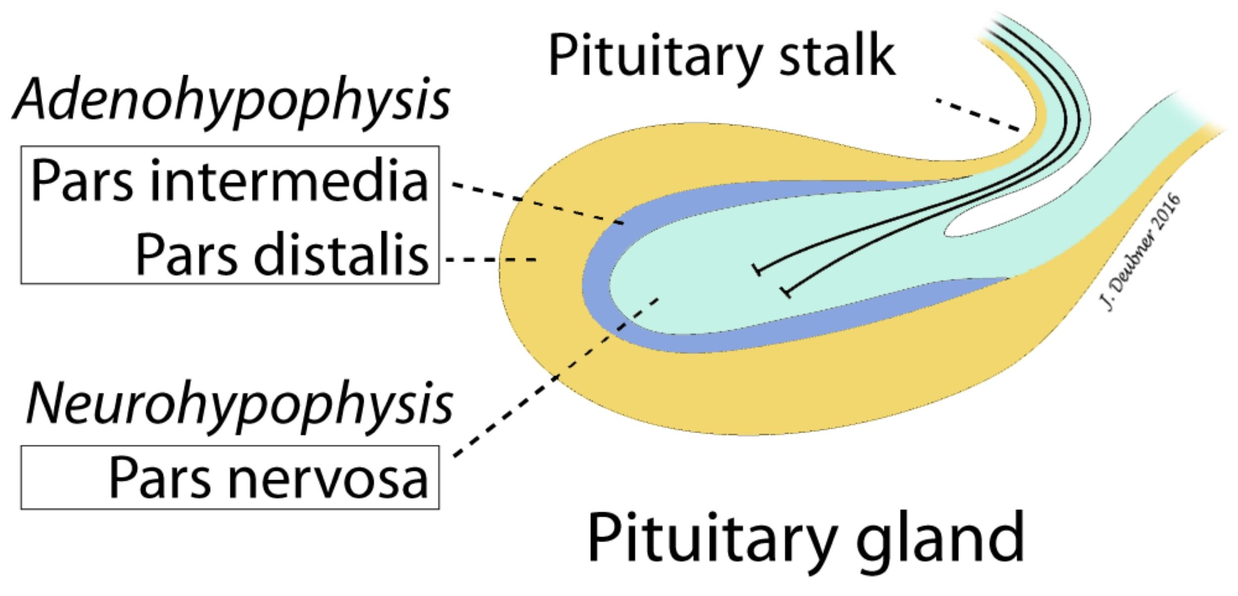
The pituitary gland (or hypophysis) comprises the adenohypophysis and the neurohypophysis (Fig. 10.3). The adenohypophysis secretes adrenocorticotropic hormone (ACTH) and TSH, along with several other hormones. The neurohypophysis secretes antidiuretic hormone (ADH, also known as vasopressin), along with other hormones. Although the hormones of the adenohypophysis are produced locally, those of the neurohypophysis are produced in the hypothalamus and are then transported to the posterior pituitary. Hormonal or nervous signals from the hypothalamus control secretion by the pituitary.

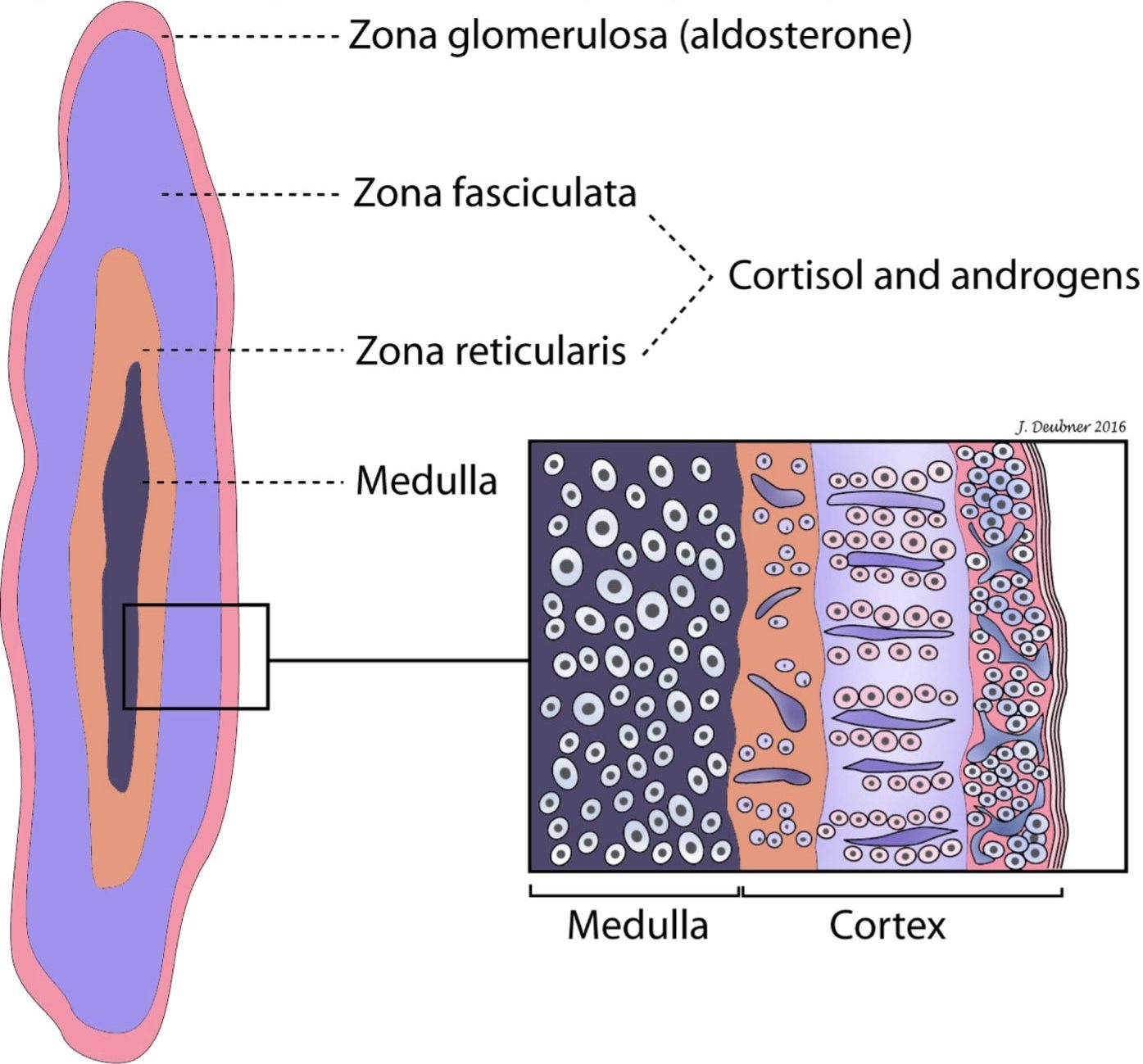
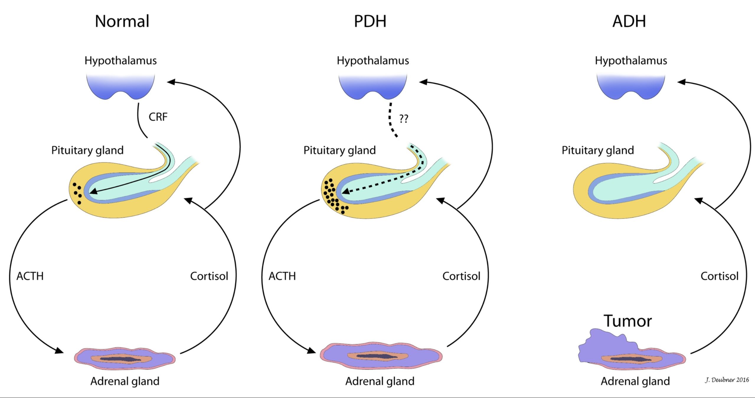
Each adrenal gland consists of a cortex and a medulla. The adrenal cortex secretes corticosteroids while the adrenal medulla secretes epinephrine and norepinephrine. In the cortex, mineralocorticoids (including aldosterone), glucocorticoids, and androgens are produced by the zona glomerulosa (ZG), zona fasciculata (ZF), and zona reticularis (ZR), respectively (Fig. 10.4). Small amounts of androgens are also produced in the ZF, and small amounts of glucocorticoids are also produced in the ZR. The secretion of mineralocorticoids is mainly controlled by ECF concentrations of angiotensin II and potassium. The secretion of glucocorticoids is controlled through the hypothalamic-pituitary axis by the action of corticotropin releasing hormone (CRH) and ACTH. As serum glucocorticoid concentrations rise, they exert a negative feedback effect on the hypothalamus and anterior pituitary to curtail ACTH secretion (Fig. 10.5).


