Pearls
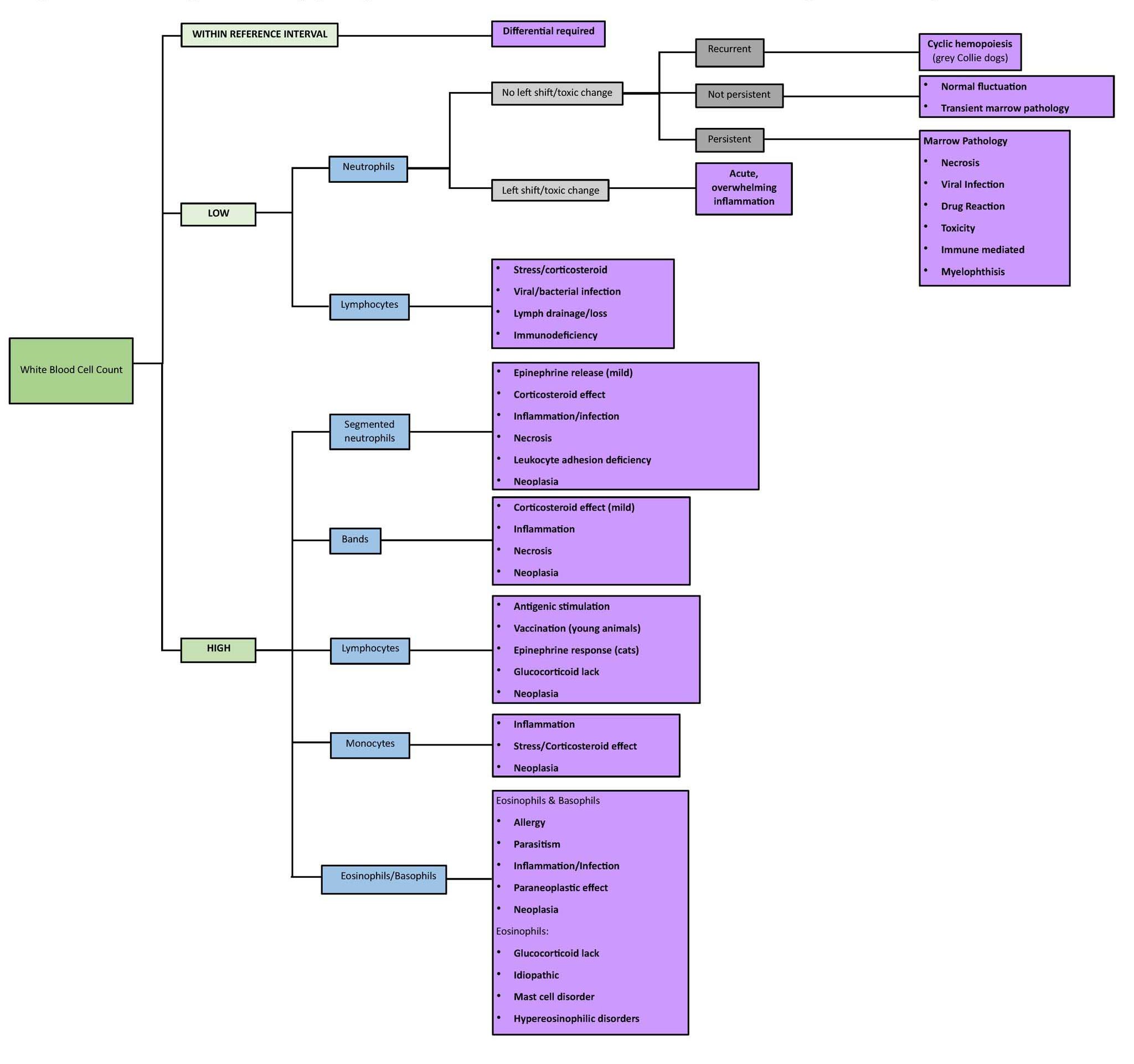
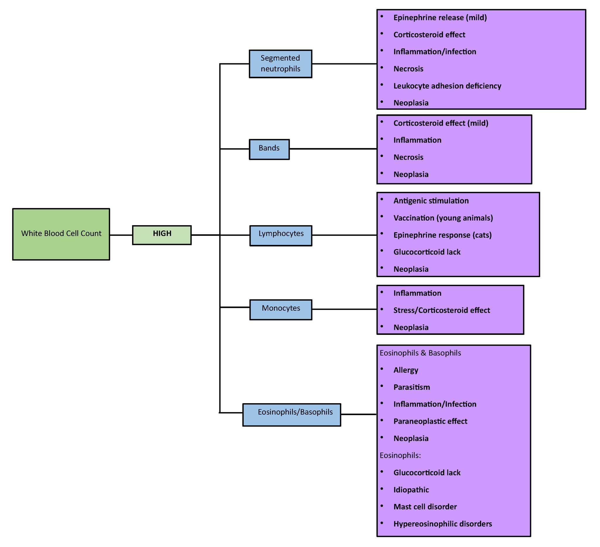
- See Fig. 2.14 for an algorithm of peripheral blood leukocyte abnormalities according to cell counts.
- When evaluating the leukogram, look at the total WBC count first to see if it is high, low, or “normal” relative to the RI.
- Then look at the ABSOLUTE numbers on the differential. The percentages are only used to calculate the absolute counts and, on their own, can be quite deceptive.
- Remember that neutrophilia is not always caused by inflammation. Excitement and stress can also result in mild neutrophilia. Interpret the leukogram as a whole and in relation to the signalment, history, and other findings to help decide the cause of neutrophilia.
- Keep in mind the effect “stress” (high levels of endogenous cortisol) or corticosteroid therapy can have on the leukogram, and that there are species differences.A “normal” differential in an extremely ill animal (especially dog) can be highly significant, suggesting hypoadrenocorticism, so always interpret the leukogram in relation to the history and clinical findings.
- DO NOT IGNORE DATA JUST BECAUSE IT IS NOT FLAGGED! “Normal” results could be highly significant, depending on the situation.
- Inflammation can be demonstrated in many different ways in the leukogram. The changes will depend on the duration, the extent (localized vs. diffuse), the cause (e.g. allergic inflammation can result in eosinophilia and/or basophilia; mycobacterial infection can result in monocytosis without neutrophilia), the ability of the bone marrow to respond (there are species differences), and treatment. For these reasons, several different patterns can be seen on the leukogram.
- Of particular concern are situations where there are high numbers of immature neutrophils (bands and possibly even earlier stages), particularly if mature neutrophil numbers are below or within the RI rather than being elevated (known as a degenerative left shift). This is interpreted as an inability of the bone marrow to adequately respond to inflammation; the inflammation is usually acute and overwhelming. The post-mitotic storage pool of neutrophils has been depleted and immature neutrophils (bands, and sometimes metamyelocytes and myelocytes) are being released. Toxic change would be expected to accompany the left shift. Immediate medical intervention is usually indicated.
- Toxic change indicates immaturity of the neutrophil cytoplasm and usually accompanies a significant left shift, meaning there are increased numbers of immature neutrophils.
- A situation where mature neutrophils are elevated with or without increases in immature cells is usually less of a medical emergency. Nevertheless, the disease process could be ultimately fatal or debilitating and the cause must be determined.
- Keep in mind that changes can occur quickly in the blood. A single CBC reflects the peripheral blood at only one point in time; histopathology of an inflammatory lesion tells a story of what has happened over a period of time. In some circumstances, it is possible to assign a duration (acute, chronic) based on leukogram changes; other times, when changes are not as dramatic, it can be impossible to make this assessment. Marked neutrophilia (again there are species differences as to what is “marked”) with or without immature neutrophils usually indicates chronicity and there is probably neutrophilic hyperplasia in the marrow. Neutropenia with increased immature cells usually indicates acute, severe inflammation; marrow stores are likely depleted.
- Do not become frustrated trying to assign a duration to inflammatory changes. Many times, it would be over interpretation of the data to attempt to do so. The important issue is to decide what the changes mean to the animal- how well is the animal responding to the process? Once more, as with the hemogram, following the leukogram over time can be extremely useful and aid in prognostication.
- Leukogram changes can signal marrow disorders such as neoplasia, injury, degenerative conditions, etc. Bone marrow examination is an extremely useful diagnostic aid, particularly if there are unexplained findings or atypical cells on peripheral blood examination. It is important that veterinarians learn how to obtain good bone marrow samples and prepare good bone marrow smears (see video on bone marrow collection).


