Myelodysplastic Syndrome
MDS is a clonal proliferative disorder in humans characterized by abnormal development and maturation of hemopoietic cells which may result in ineffective hemopoiesis and peripheral blood cytopenias. MDS has been reported in FeLV-infected cats and has been shown to be a clonal disorder under these circumstances. In dogs and horses, clonality is suspected, though not proven. MDS-related cytopenia(s) can manifest as anemia, thrombocytopenia (resulting in primary hemostatic defects), and neutropenia (resulting in increased susceptibility to infections). Bone marrow is usually hypercellular, however, dyshemopoiesis results in impaired maturation of affected cell lines and abnormal morphologic findings. The latter include fragmentation of nuclei, asynchronous maturation of nuclei relative to cytoplasms, abnormal lobulation of megakaryocyte nuclei, increased apoptosis of hemopoietic cells, and hypersegmentation of granulocyte nuclei. MDS is considered a pre-leukemic disorder as it may progress to acute leukemia. However, the clonal disorder MDS, must be differentiated from non-neoplastic conditions, such as nutritional deficiencies, congenital defects, and drug and toxin exposure, that can also be associated with maturation defects in hemopoietic cells. Increased numbers of blast cells, up to 30% myeloblasts or rubriblasts, along with other abnormal morphologic features, can be useful in differentiating the neoplastic and non-neoplastic disorders.
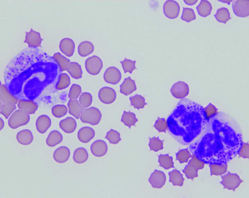
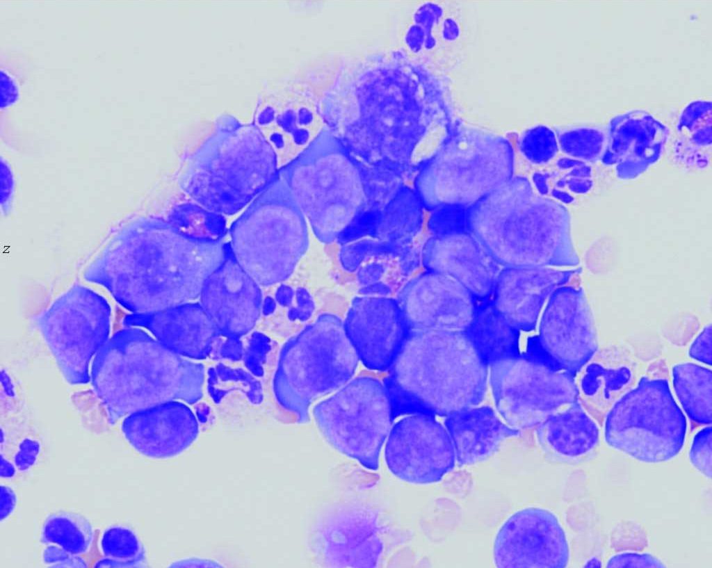
See Fig. 3.10 for a rare case of basophilic leukemia in a cat. Also see Fig 3.11 for thoracic fluid from a dog with multicentric lymphosarcoma.


Process of producing all the cells found in the blood, including RBCs, WBCs, and platelets.
Decrease in the number of neutrophils in peripheral blood below the reference interval for the species.

