Mast Cell Neoplasms
Although mast cells are bone marrow-derived hemopoietic cells, they are not seen in the peripheral blood under normal circumstances and they are also not included in the classification schemes for myeloid neoplasms. However, mast cell tumors are important neoplasms in veterinary medicine and are seen in cats and dogs, and less commonly in other domestic species.
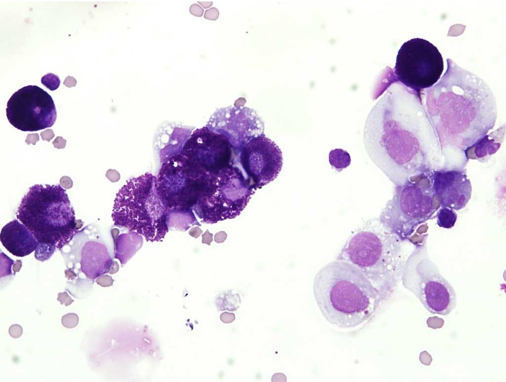
Most mast cell tumors are cutaneous and not associated with systemic disease. However, disseminated mast cell sarcoma is sometimes seen. Bone marrow, spleen, liver, and lymph nodes may be sites of neoplastic proliferation, with or without cutaneous involvement. Cats may be presented with abdominal enlargement, often accompanied by vomiting. A markedly enlarged spleen may be palpated. Fine needle aspiration of the spleen or retrieval of abdominal fluid is diagnostically useful (Fig. 3.9). Animals with systemic mast cell neoplasia may be presented with vomiting and intestinal hemorrhage. Malignant mast cells sometimes release histamine which stimulates H2 receptors in the stomach and duodenum, resulting in increased acid release, mucosal ulceration, hemorrhage, and vomiting.

Malignant mast cells may be released into the peripheral blood when bone marrow is involved. Morphologically, these cells are often well-differentiated and granulated, despite their malignancy. Mast cells are generally larger than peripheral blood leukocytes and are often swept to the feathered edge of the peripheral blood smear. A buffy coat preparation of the peripheral blood may be useful to rule in or out the presence of circulating mast cells.
Mast cells can appear in the peripheral blood with both neoplastic and non-neoplastic/inflammatory conditions. Therefore, caution must be exercised when diagnosing mast cell neoplasia based solely on their presence in the peripheral blood. Mastocytemia is a term used to describe the presence of mast cells in the peripheral blood, and is applied to both neoplastic and non-neoplastic disorders. The number of circulating mast cells with inflammatory processes is generally low. Potential causes of non-neoplastic or reactive mastocytemia include, but are not limited to: parvoviral enteritis, cutaneous hypersensitivities and parasitic infections, IMHA, bacterial peritonitis, pancreatic necrosis, and pleuritis. Neoplastic mastocytemia can occur as a progression of systemic mast cell neoplasia which usually begins at a visceral or cutaneous site. Neoplastic mastocytemia can also be due to a primary bone marrow-derived mast cell leukemia. Integration of all the clinical and laboratory information is important, as always, to distinguish between neoplastic and non-neoplastic conditions, as well as mastocytemia due to primary bone marrow neoplasia versus extension from a solid tissue mast cell tumor.
Edge of the blood smear farthest from where the droplet of blood is placed.
Smear made from a concentrated preparation of peripheral blood leukocytes.

