Granulopoiesis
A single common myeloid progenitor differentiates into all of the hemopoietic cell lines, with the exception of lymphocytes (Fig. 1.1, Chapter 1. Erythrocytes). The bone marrow is the most effective and common site for hemopoiesis, including leukocyte production, in domestic animals. However, extramedullary production can also occur in other organs, such as the spleen and liver.
The production of granulocytes from bone marrow stem cells through to mature neutrophils, eosinophils, and basophils is known as granulopoiesis. Granulocytes are mobile leukocytes with the ability to phagocytose, degrade, and/or kill microorganisms, depending on the offending agent and granulocyte lineage.The commitment to neutrophilic, eosinophilic, or basophilic leukocytes occurs at an early level of common myeloid progenitor differentiation and is dependent on the presence and relative concentrations of several growth factors and cytokines. The most important of these are granulocyte-colony stimulating factor (G-CSF) for neutrophils; interleukin(IL)-5 for eosinophils; and stem cell factor (SCF) for basophils.
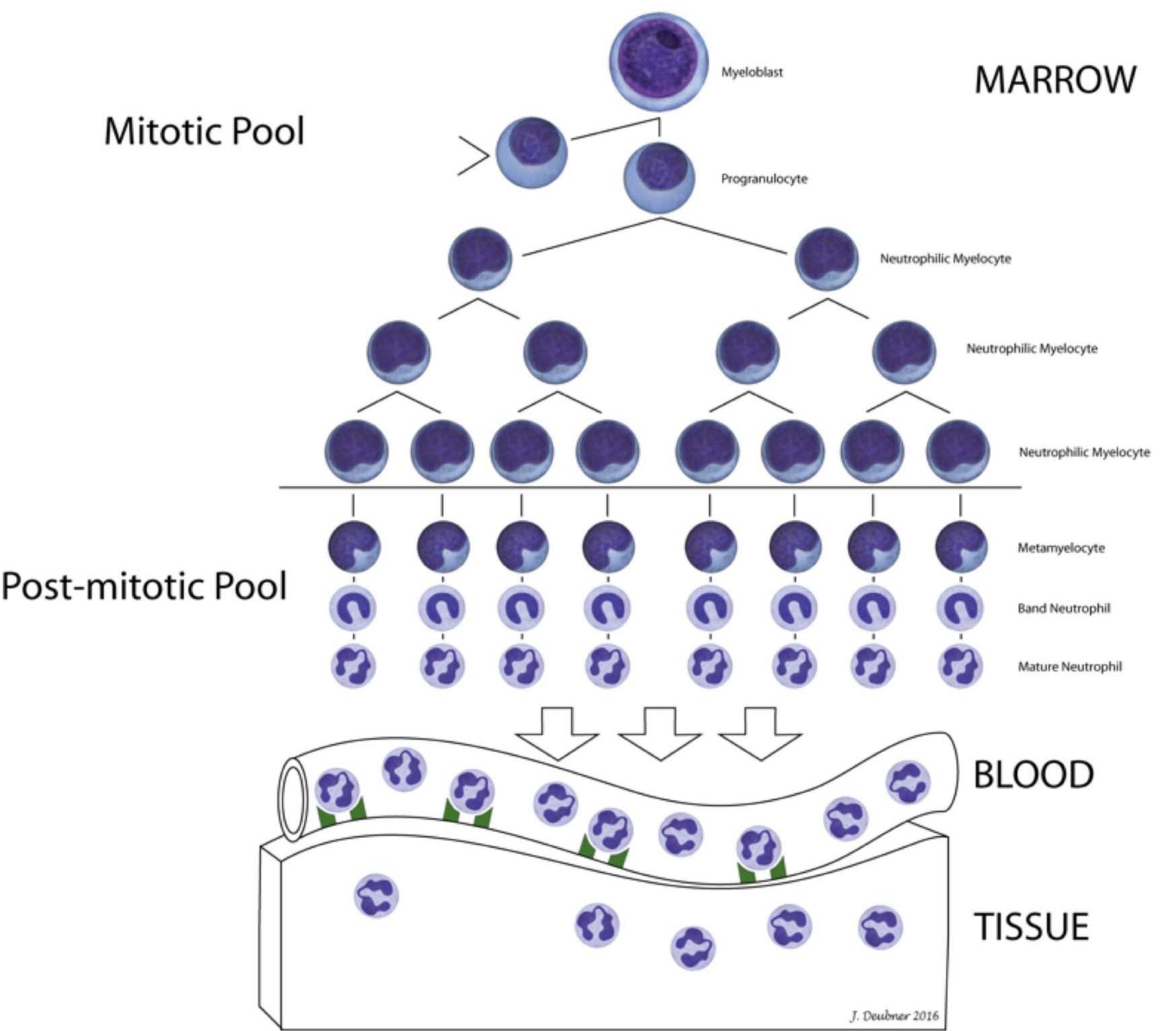
The earliest committed granulocyte precursor that can be recognized by light microscopy is the myeloblast (see Chapter 3. Hemopoietic Neoplasia, Fig. 3.8). Myeloblasts are agranular primitive cells which differentiate to promyelocytes with the synthesis of primary (azurophilic) granules. Primary granule contents include myeloperoxidase, lysozyme, and defensins. Synthesis of secondary (specific) granules defines the developing granulocyte as a neutrophilic, eosinophilic, or basophilic myelocyte. Secondary neutrophilic granule contents include lysozyme, lactoferrin, and gelatinase which, together with cytoplasmic organelles, enable phagocytosis and killing of microbes. Up to five mitotic divisions occur from myeloblast to myelocyte stages, and the total time in the mitotic (proliferating) phase of granulopoiesis is about 3 days. The post-mitotic (maturation and storage) phase comprises metamyelocytes, bands, and mature granulocytes. The interval from metamyelocyte to mature segmented granulocyte is also about 3 days for most animals (Fig. 2.1).

Can have different hematologic meanings: 1. referring to all hemopoietic cells in the bone marrow except lymphocytes, 2. referring to all bone marrow leukocytes except lymphocytes.
Process of producing all the cells found in the blood, including RBCs, WBCs, and platelets.
Part of hemopoiesis dealing with the production of granulocytes from stem cells to mature circulating neutrophils, eosinophils, and basophils.
White blood cell (WBC); includes neutrophils, eosinophils, basophils, monocytes, lymphocytes, mast cells.
Granulocyte-Colony Stimulating Factor; important growth factor in early granulopoiesis.
Stage in granulopoiesis following the committed stem cell stage; earliest form recognizable as a granulocyte precursor.
Stage in granulopoiesis following myeloblast; synthesis of primary (azurophilic) granules occurs.
Granules containing myeloperoxidase, lysozyme, and defensins; found in promyelocytes.
Stage in granulopoiesis following promyelocyte; synthesis of secondary (specific) granules occurs. Type of granulocyte can now be determined (neutrophil, eosinophil, or basophil).
Stage in granulopoiesis following myelocyte; nucleus begins to become indented.
Immature neutrophil with an unsegmented (band-shaped) nucleus

