Erythrocyte Values
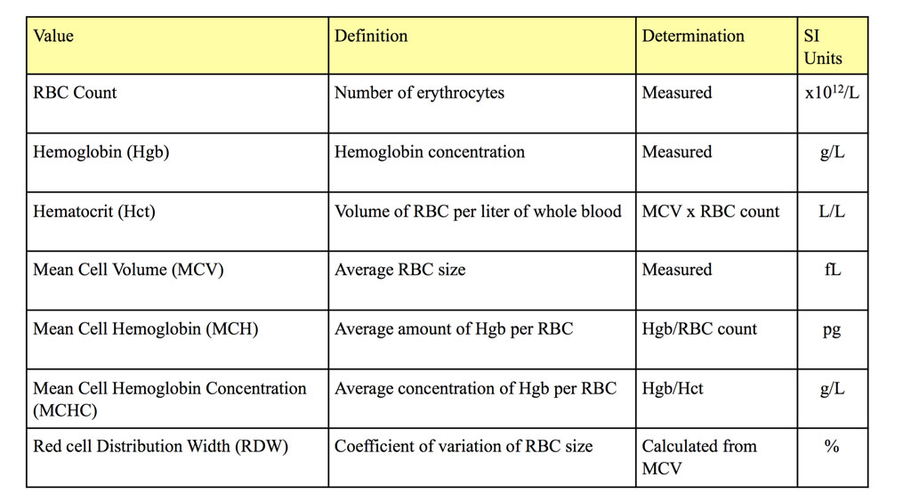
The erythron refers to the total mass of circulating RBCs and their precursors in the bone marrow, whereas the erythrogram evaluates several measurements and descriptive qualities of only RBCs in the peripheral circulation. The erythrogram is that part of the complete blood count (CBC) that describes erythrocyte morphology, number, size, and hemoglobin content determined by both peripheral blood smear examination and a combination of measurements and calculations, usually with the aid of automated instruments. These erythrocyte values include: RBC count, hemoglobin (Hgb), hematocrit (Hct), packed cell volume (PCV), mean cell volume (MCV), mean cell hemoglobin (MCH), mean cell hemoglobin concentration (MCHC), red cell distribution width (RDW), reticulocyte percent and absolute count, and reticulocyte production index (RPI) (dogs only). Most automated instruments provide an RBC count, Hgb, and MCV, which are measured, and Hct, MCH, MCHC, and RDW, which are calculated (Table 1.2). The RPI is calculated using the reticulocyte percent, which is usually determined manually. A PCV is determined manually using a microhematocrit tube and centrifuge and is performed when the Hct cannot be determined using an automated instrument for several reasons, including RBC agglutination.

- RBC: The RBC count is the number (N) of erythrocytes, usually written as N x 1012 per liter (/L).
- Hgb: Hemoglobin concentration is measured and reported as grams per liter (g/L).
- MCV: Automated instruments measure the sizes of several thousand red cells and then report the average of these sizes as the MCV in femtoliters (1fL = 10-15L).
- Hct: The Hct, calculated by multiplying the MCV by the RBC count, is the volume of red cells per liter of whole blood. For example, if the MCV is 72.4 fL and the RBC count is 4.77 x 10¹²/L, the Hct is (72.4 x 10⁻¹⁵L) X (4.77 x 10¹²/L) = 0.345 L/L.
- PCV: The Hct is equivalent to the PCV, but the latter term is generally reserved for those times when a small tube of blood (microhematocrit tube) is centrifuged and the volume of packed RBCs relative to the total volume of the sample is reported as a percentage. PCV may be reported, for example, when RBC agglutination precludes obtaining many erythrocyte values using an automated instrument.
- MCH: The MCH is the average amount of hemoglobin per red cell and is calculated by dividing the Hgb by the RBC count. For example, if the Hgb is 115 g/L and the RBC count is 4.77 x 10¹²/L, the MCH is 115 g/L divided by 4.77 x 10¹²/L = 24.1 x 10⁻¹²g or 24.1 picograms (pg).
- MCHC: The MCHC is the average concentration of hemoglobin per erythrocyte and is calculated by dividing the Hgb by the Hct. For example, if the Hgb is 115 g/L and the Hct is 0.345 L/L, then the MCHC is 115 g/L divided by 0.345 L/L = 333 g/L.
- RDW: The red cell distribution width is calculated from the MCV and describes the coefficient of variation of the red cell sizes. The formula for RDW is the standard deviation of the MCV divided by the MCV. The RDW, in principle, correlates with the degree of RBC anisocytosis observed on blood smear evaluation.
- Reticulocyte count: When anemia is detected, a reticulocyte count is done to evaluate the bone marrow response and differentiate regenerative from nonregenerative anemia. Peripheral blood is stained, usually with NMB, so that residual ribosomes, mitochondria, and other cytoplasmic organelles are aggregated and precipitated in strands or clumps in immature erythrocytes. Although these cells are equivalent to polychromatophilic cells with Romanowsky stains, they are more easily visualized and enumerated with NMB staining. The number of reticulocytes is expressed as a percentage. The percentage of reticulocytes can be multiplied by the RBC count to determine the absolute number of reticulocytes in a volume of blood. This calculation adjusts for the higher relative percentage of reticulocytes when mixed with fewer mature erythrocytes in anemia.
Additional Notes
Occasionally polychromasia is present without an accompanying anemia. A reticulocyte count is also performed in this situation to quantify what may represent recovery from prior or ongoing low grade hemorrhage or hemolysis.
In addition to aggregate reticulocytes (with precipitated clumps or strands of RNA, Fig. 1.4), punctate reticulocytes (with small blue dots of precipitated RNA) are also seen, particularly in cats. These are not enumerated when doing a reticulocyte count as they represent aged aggregate reticulocytes and do not reflect current regeneration (Fig. 1.5).
Reticulocyte counts are not routinely performed in anemic horses as this species rarely releases immature RBCs into the peripheral circulation, even with regenerative anemias.
- RPI: The RPI is sometimes reported for canine samples and is a calculation designed to correct the reticulocyte count for the severity of the anemia and for the longer maturation period for early-released reticulocytes. The RPI has been adapted from human medicine and requires that a set value be assigned for the normal hematocrit, which is not always correct for the individual animal. The various manipulations of the reticulocyte percent are designed to determine if the regenerative response is adequate for the degree of anemia.
All circulating erythrocytes and erythrocyte precursors within the body.
All tests on the complete blood count (CBC) that evaluate erythrocytes, including morphologic examination on the peripheral blood smear.
Number of erythrocytes per liter of whole blood (measured).
Volume of erythrocytes per liter of whole blood. Reported as L/L (calculated: MCV x RBC count). Equivalent to PCV (%) determined by centrifugation of blood in a microhematocrit tube
Equivalent to the hematocrit, but measured using a microhematocrit tube.
Average erythrocyte size in femtoliters (measured or calculated PCV or Hct÷RBC).
Average amount of hemoglobin per erythrocyte (calculated: hemoglobin÷RBC count).
Average concentration of hemoglobin per erythrocyte (calculated: hemoglobin÷hematocrit).
Red cell distribution width; a measure of RBC variation in size (anisocytosis) within a blood sample (calculated)
Reticulocyte production index; a calculated value designed to determine if the regenerative response is adequate for the degree of anemia. Corrects for the longer life of a young reticulocyte and the relative increase in reticulocytes in the presence of a low hematocrit (anemia). Reported for dogs only.
Clumping of erythrocytes due to antibody interactions. Must be differentiated from rouleaux formation.
Variation in cell size.
Anemia in which the bone marrow does not respond by producing more RBCs; there are many reasons why this may occur including bone marrow injury, chronic renal and inflammatory disease.
Anemia in which the bone marrow responds appropriately by increasing the production of RBCs; polychromasia (except horses) and often macrocytosis are seen in peripheral blood.

