Epithelial Tumors
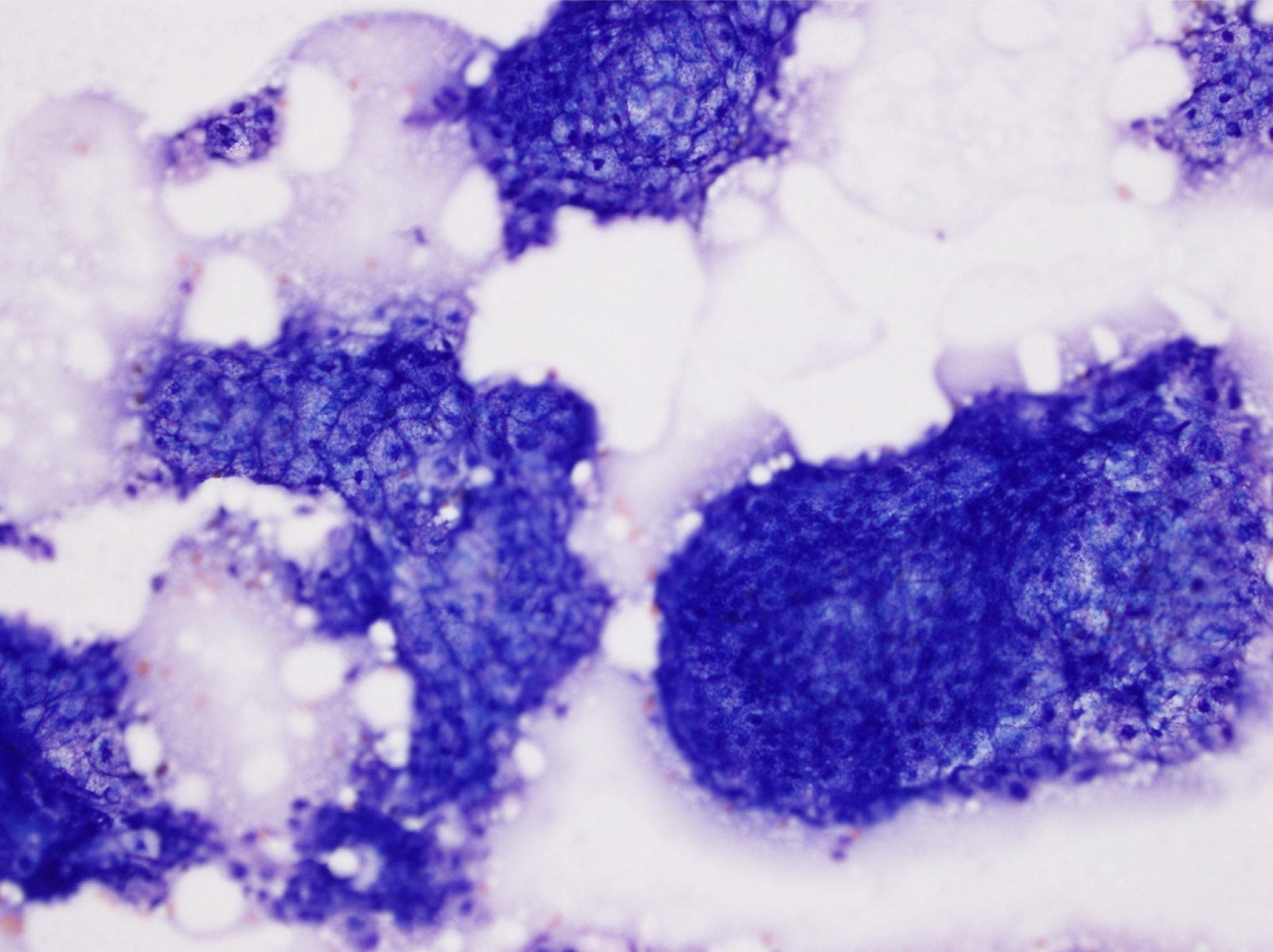
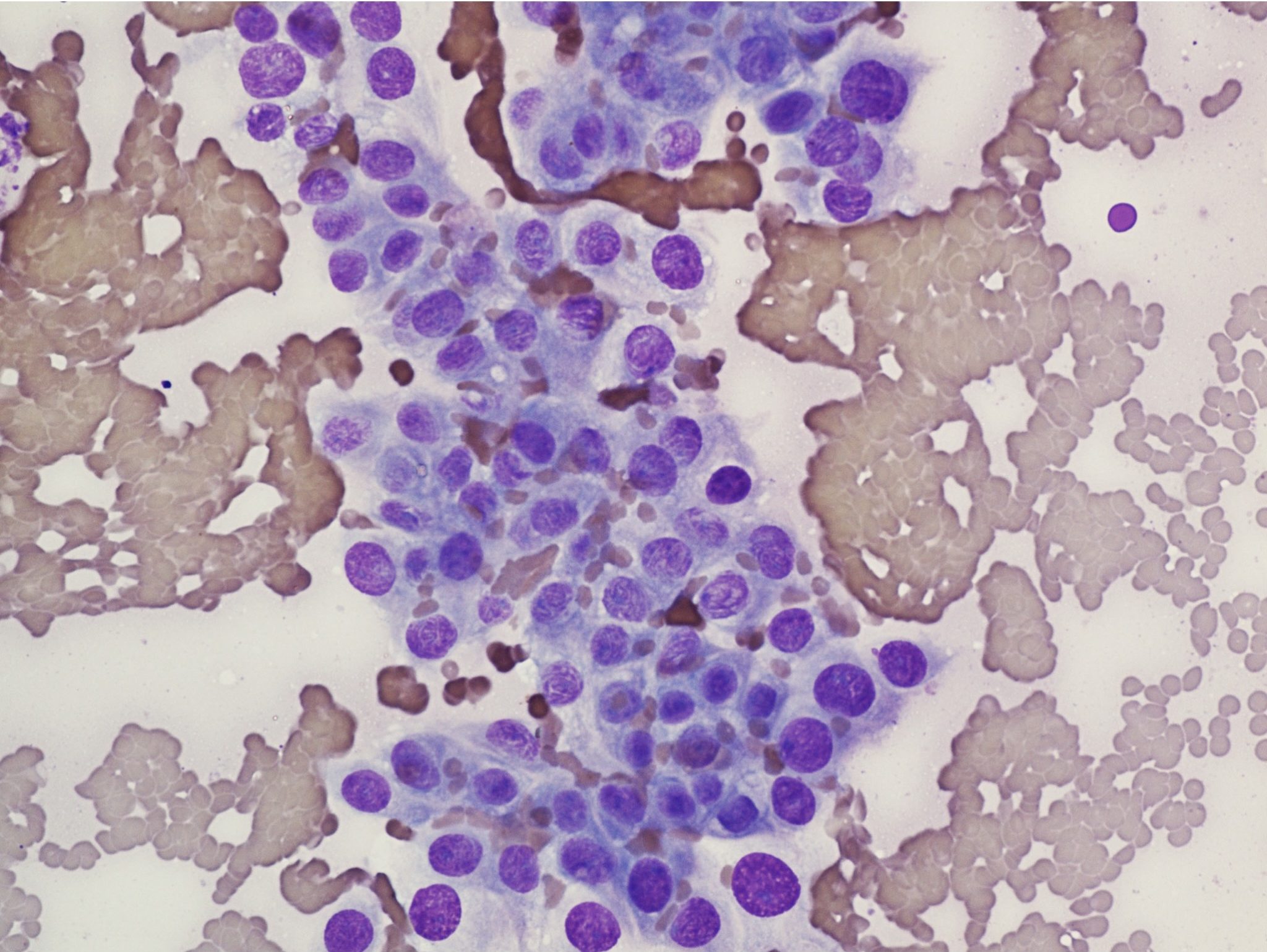
Epithelial tumors tend to exfoliate well and cells are often in clumps or sheets due to cell-cell attachments. Epithelial cells are usually round to polyhedral with distinct cell borders, round nuclei, and abundant cytoplasm (i.e. a low nuclear to cytoplasmic ratio). However, exceptions do exist, for example, basal cells have a high nuclear to cytoplasmic ratio due to the presence of scant cytoplasm. Benign epithelial cell tumors closely resemble the normal tissue and, sometimes, the diagnosis of a benign tumor can only be made due to knowledge that a mass exists. Sheets or clumps of benign epithelial cells are well organized, and individual cells resemble one another in size, shape, staining characteristics, and nuclear to cytoplasmic ratio. If the epithelial tissue is glandular in origin, cells may form rosettes, acini, or ducts or display secretory activity. In general, benign epithelial tumors are called adenomas, for example, sebaceous adenoma; malignant epithelial tumors are called carcinomas, for example, squamous cell carcinoma. See Fig. 5.12 for benign (well differentiated) epithelial cells and Fig. 5.13 for malignant (poorly differentiated) epithelial cells.


Comparison of the relative size of the nucleus and cytoplasm of a cell; a cell with abundant cytoplasm has a low N:C ratio, while a cell with a large nucleus and scant cytoplasm has a high N:C ratio. A high N:C ratio may be seen in neoplastic cells, but also in some normal cells (e.g. small lymphocyte).
Malignant neoplasm of epithelial origin, e.g. squamous cell carcinoma.

