Eosinophils
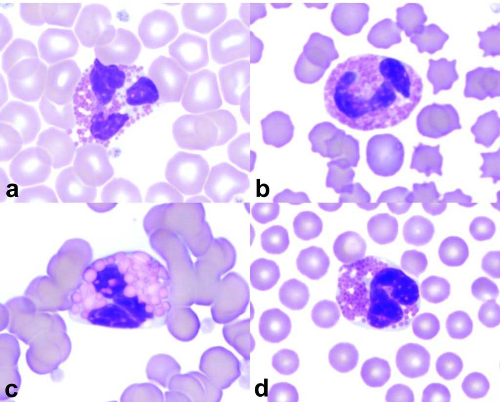
Eosinophils are derived from the common myeloid progenitor along with basophils, and parallel neutrophils in development and maturation. GM-CSF, IL-3, and IL-5, produced by T-helper lymphocytes, are required for eosinophil differentiation. IL-5, in particular, is required for eosinophil maturation and function. Secondary granules distinguish this granulocyte as an eosinophil at the myelocyte stage in the bone marrow. The size, shape, and number of granules vary with the species, but most are pink to orange and very distinctive with Romanowsky stains (Fig 2.8). The granules contain proteins and enzymes, namely, major basic protein, eosinophil peroxidase, eosinophil cationic protein, and eosinophil-derived neurotoxin. Eosinophil granules enable anthelminthic activities, suppression of immediate hypersensitivity reactions, promotion of inflammation in certain other allergic diseases, phagocytosis of some microbes and proteinaceous and cellular material, tumor cell killing, and immune reactions involving T lymphocytes.

Eosinophils circulate in the peripheral blood for a very short time (about 1 hour in the dog), roll along the endothelium, and migrate into tissues in a manner similar to neutrophils. Eosinophils reside in skin and submucosal tissues where they guard against various pathogens from the external environment. Peripheral blood eosinophil numbers are often a poor reflection of tissue concentrations, and significant eosinophilic inflammation can be present in tissue sites without evidence on the CBC.
Eosinophilia
Elevated eosinophil numbers on the CBC warrant investigation for parasitic infections, hypersensitivity disorders, inflammatory conditions involving primarily eosinophil recruitment or infiltration (of which there are many types involving many tissues), eosinophilic leukemia, lymphoid neoplasia, mast cell disorders, including neoplasia, and hypoadrenocorticism. Eosinophils act together with T lymphocytes and release toxic mediators onto the surface of parasites. In hypersensitivity reactions, eosinophils are recruited to neutralize inflammatory mediators released from basophils and mast cells, and to carry out other immunosuppressive activities. Although primary eosinophilic neoplasms are rare, eosinophilia can accompany lymphosarcoma and mast cell tumors. Presumably, the neoplastic cell population elaborates mediators that are chemotactic for eosinophils and decrease apoptosis of eosinophils in tissues. Glucocorticoid release, as expected with illness and stress, suppresses eosinophil numbers. The mechanism of glucocorticoid-induced eosinopenia is not well understood, but relates to suppression of eosinophil survival factors (GM-CSF, IL-3, and IL-5) and activation of eosinophil apoptosis. If the eosinophil count is near the upper limit of the RI or elevated in an obviously ill animal (usually a dog), lack of glucocorticoid influence should be suspected. Hypoadrenocorticism is a consideration under these circumstances.
Eosinopenia
Eosinopenia cannot be detected in many species since the lower limit of the RI may by zero or near zero. There are no known health risks associated with eosinopenia as there are with neutropenia, and low numbers can be a consequence of illness and stress which is present in many of our veterinary patients.
Granules specific to either neutrophils, eosinophils, or basophils, and containing substances used by the cell in host defence.
Referring to lymphocytes and tissues where lymphocytes develop.
Decrease in the number of eosinophils in peripheral blood. May not be detectable given the reference intervals.

