Diagnostic Washes
For certain sites, such as the airways, alveoli, nasal passages, urinary bladder, and prostate, flushing sterile isotonic fluid into the site may be necessary to dislodge cells and produce a diagnostic sample. There is no value in quantitating the nucleated cell number or protein content in such a sample because of the variable dilutional effect of the wash. Cytospin preparation to concentrate the cells is particularly useful for these often poorly cellular fluids.
Tracheobronchial Wash & Bronchoalveolar Lavage
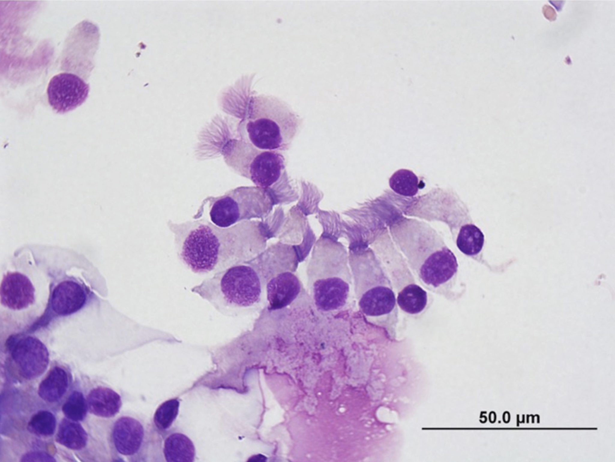
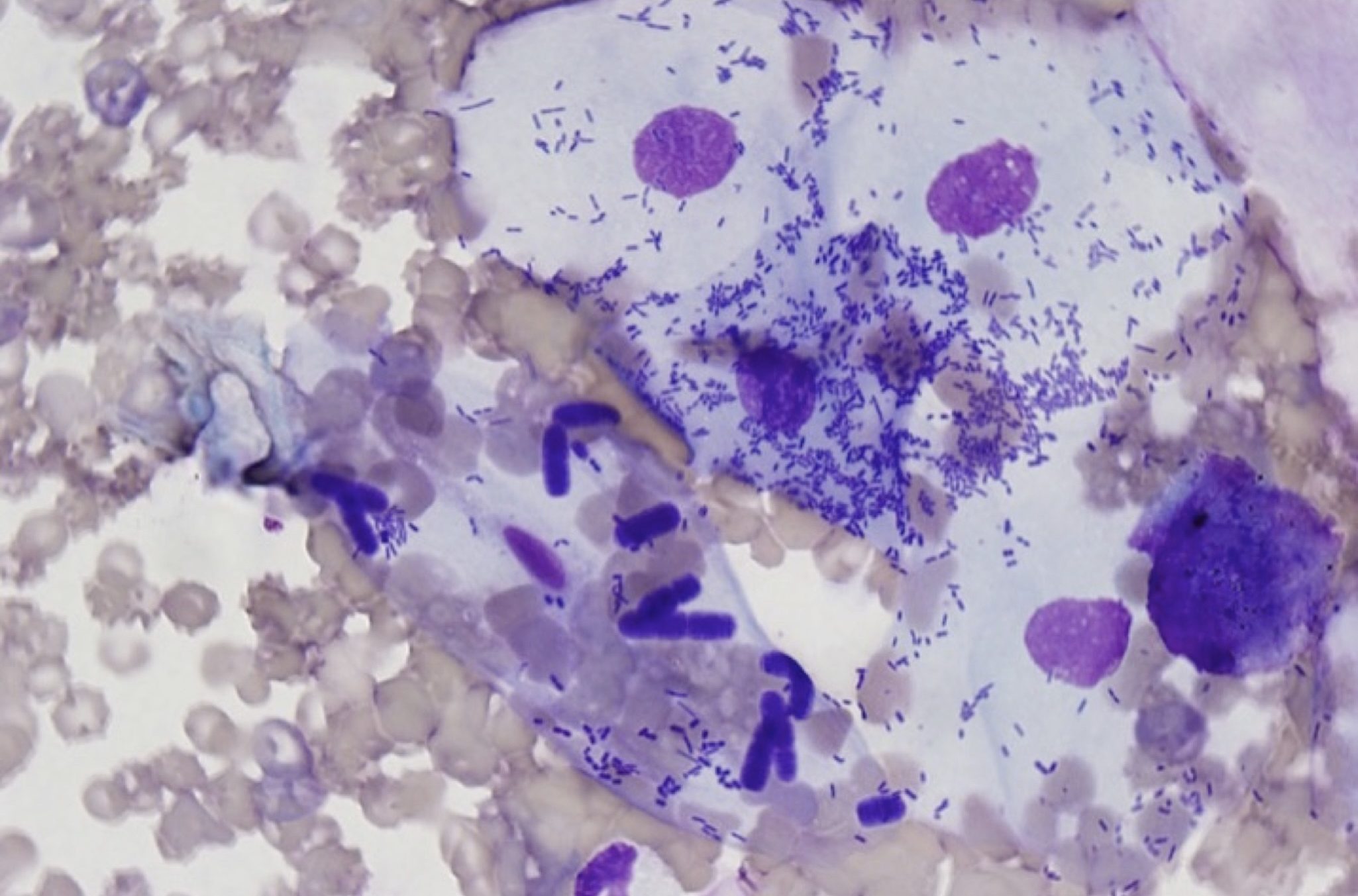
The tracheobronchial wash is valuable in assessing the airways, while bronchoalveolar lavage (BAL) is used primarily to assess the alveoli. A normal tracheobronchial wash contains ciliated columnar respiratory epithelial cells (Fig. 5.41), occasional macrophages, and little else. Samples that have been contaminated with material from the oropharynx contain a mixture of extracellular bacteria (particularly Simonsiella sp. in small animals and horses), squamous epithelial cells, and debris (Fig. 5.42). The procedure should be repeated if contamination has occurred, and bacterial culture of this material is of no value. With tracheitis, tracheobronchitis, and pneumonia, inflammatory cell numbers are increased, the type of cell varying with the cause. Allergic airway disease, for example asthma in cats or dogs, results in increased proportions of eosinophils. Fungal infection, such as blastomycosis, causes a mixed neutrophilic and macrophagic reaction; neutrophils often surround the fungal organisms, whereas, macrophages and giant cells may phagocytose organisms. Most commonly, the presence of inflammatory cells necessitates a careful search for offending organisms. Neutrophilic inflammation, especially if degenerate neutrophils predominate, suggests a bacterial cause. Although uncommon, a tracheobronchial wash can yield a neoplastic population of cells possibly admixed with inflammatory cells.


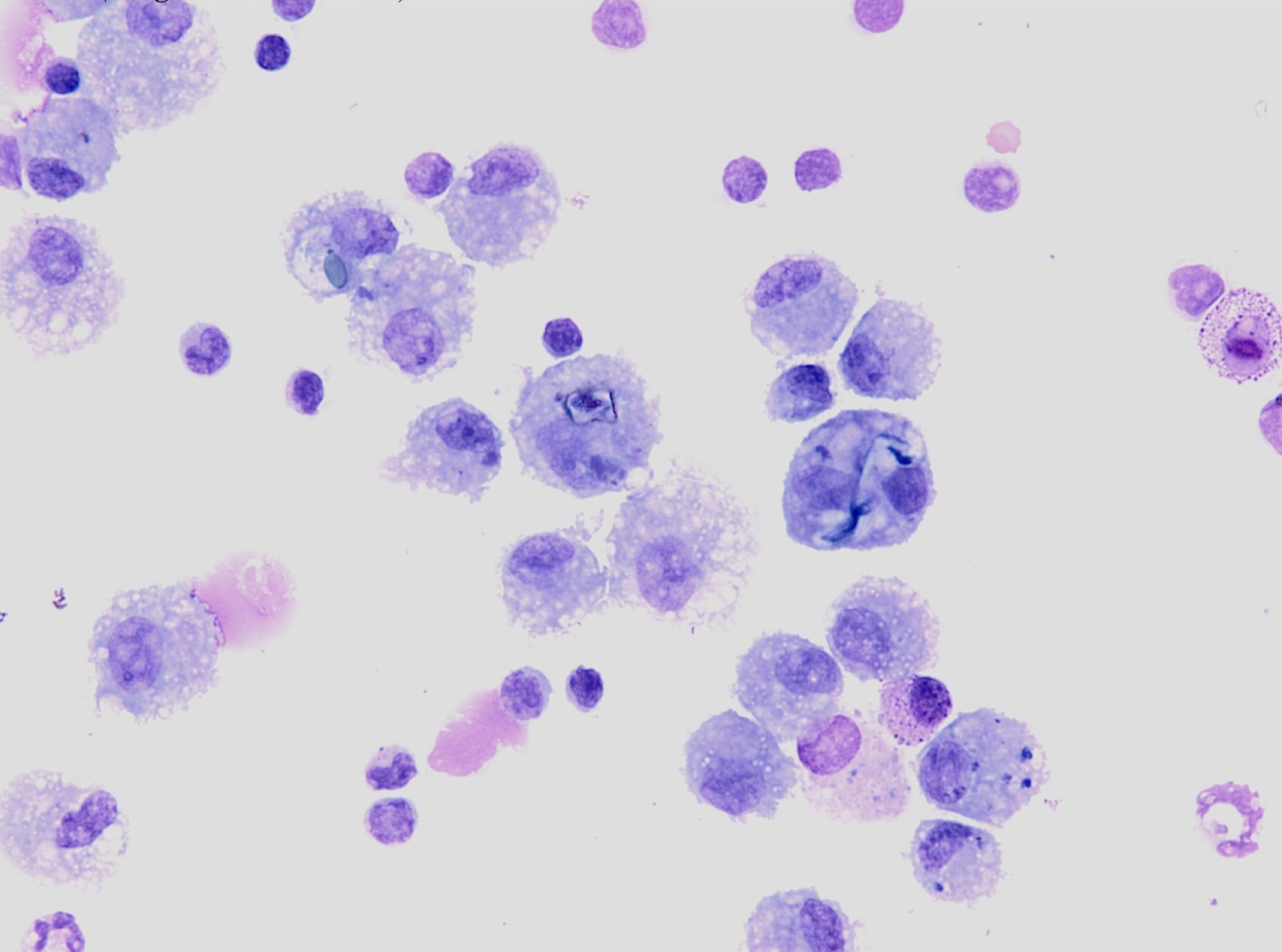
A normal BAL sample contains low numbers of alveolar macrophages with fewer small lymphocytes and occasional nondegenerate neutrophils. Cell counts can provide useful information if a constant volume of saline is infused. Macrophages may contain phagocytosed pollen granules and other particulate debris in animals kept in dusty environments (for example, horses housed in barns; Fig. 5.43). Recurrent airway obstruction (previously called heaves or chronic obstructive pulmonary disease) in horses produces a highly cellular sample comprising nondegenerate neutrophils, macrophages, and often, abundant mucus. The presence of even low numbers of mast cells and eosinophils can reflect inflammatory airway disease in horses. Horses with exercise-induced pulmonary hemorrhage often bleed into the lower respiratory tract and numerous erythrophagocytic and hemosiderin-laden macrophages are found in BAL samples.

Genitourinary Tract Wash
Prostatic or urinary bladder flushing or washing is sometimes used to obtain sufficient cells to differentiate inflammatory, neoplastic, metaplastic, and dysplastic processes, for example, if suspicious cells are seen in the urine sediment. The prostate or urinary bladder wall can also be sampled directly by fine needle with ultrasound guidance or via traumatic catheterization. These procedures result in the collection of cells that are potentially more intact and, therefore, morphologically superior to those sloughed naturally in urine. However, some clinicians may be reluctant to aspirate possibly neoplastic bladder masses due to the risk of seeding the tumor in the abdomen. Urinary tract inflammation and infection often accompany carcinoma of the urinary tract, and re-evaluation after treating the infection may be necessary along with tests such as ultrasonography and endoscopy. Also, chronic urinary tract inflammation/infection can cause epithelial dysplasia, again necessitating monitoring and additional testing in order to differentiate neoplasia and dysplasia.
Process of using a saline flush to obtain a sample of the cells within the trachea and proximal airways; may be transtracheal or endotracheal.
Process of using a saline flush delivered by endoscope to obtain a sample of the cells within the alveoli and terminal bronchioles.
Abnormal growth and maturation of the epithelium, e.g. due to chronic irritation, that may be difficult to differentiate from neoplastic changes.

