Criteria of Malignancy
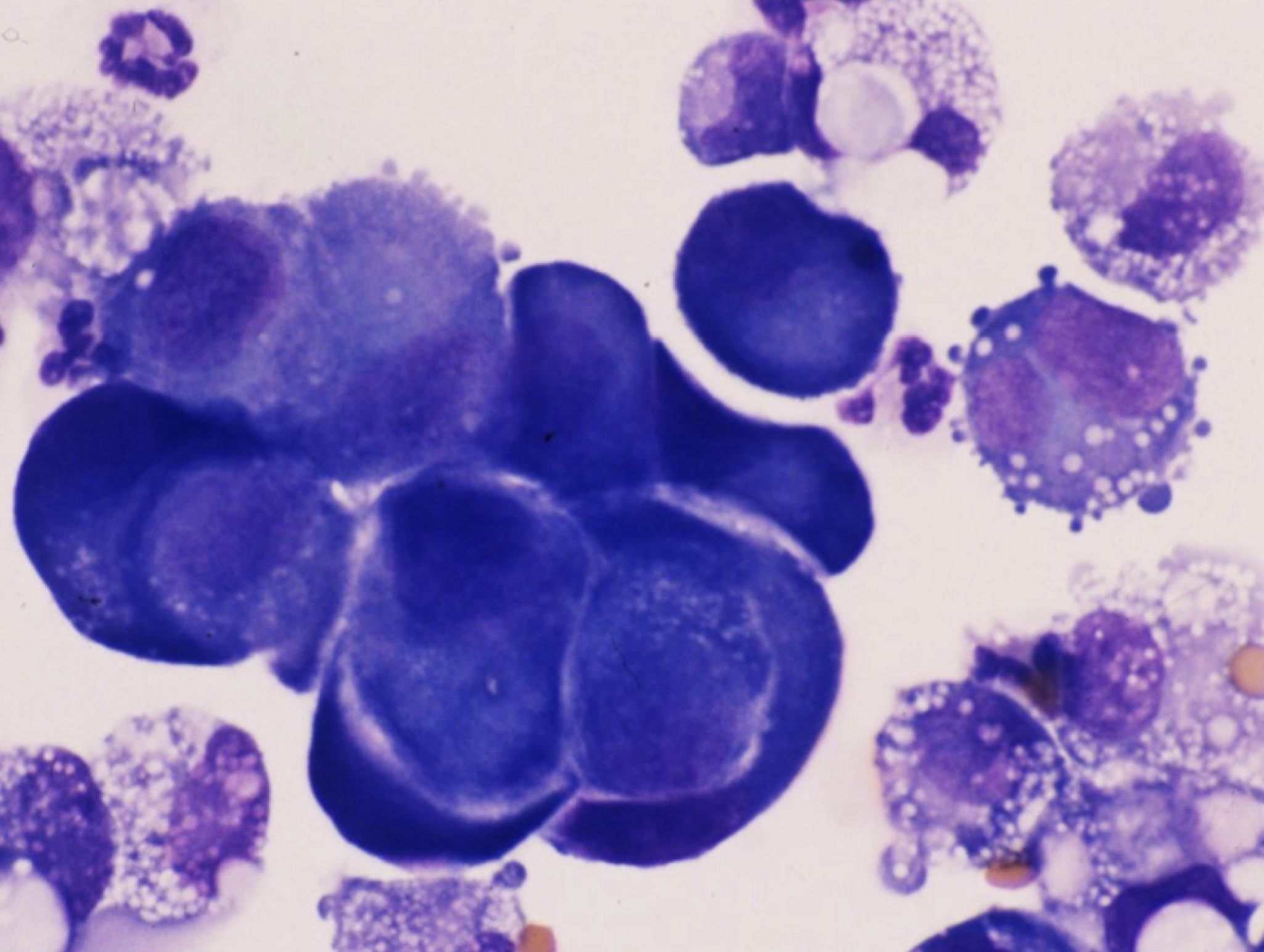
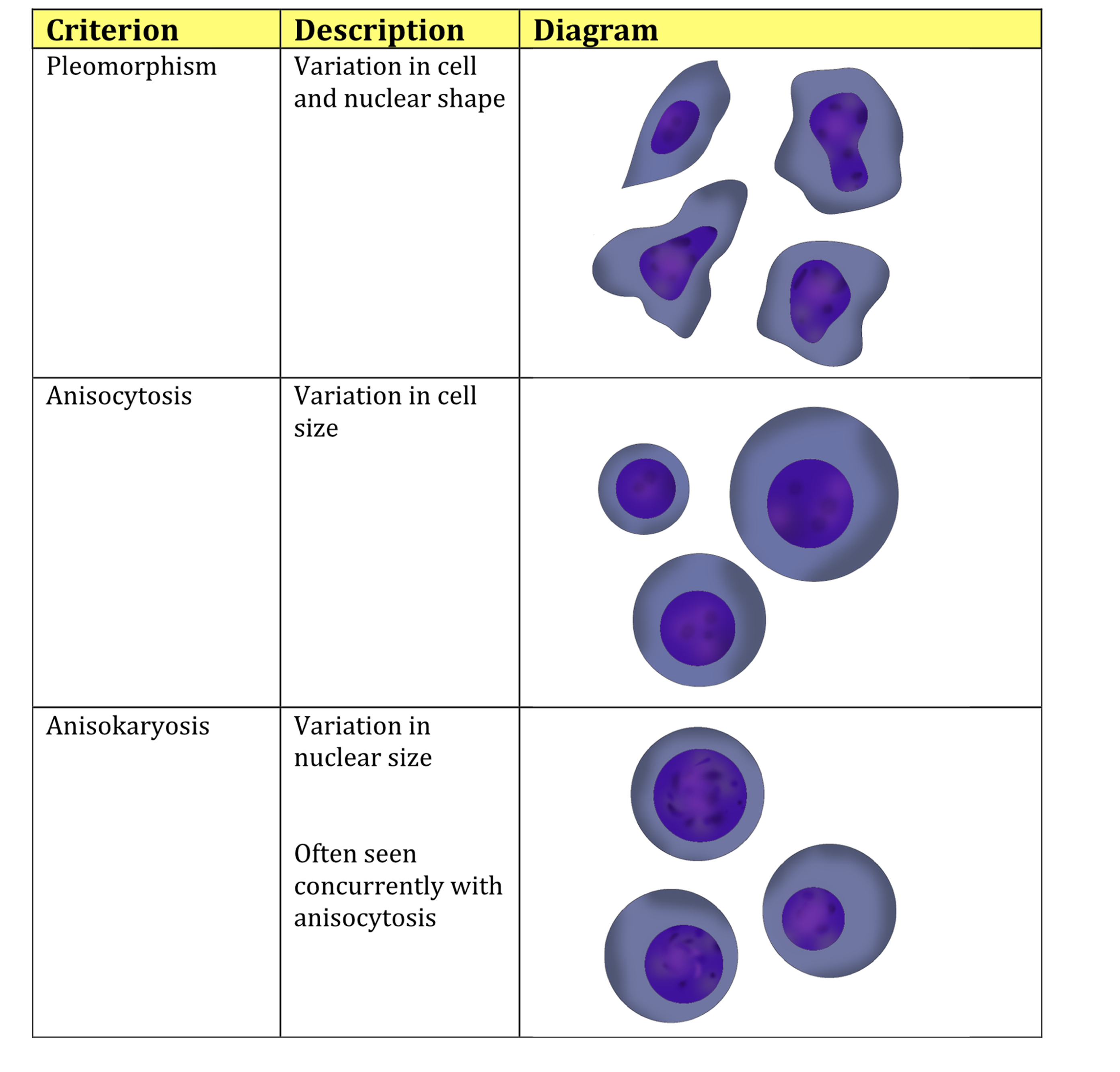
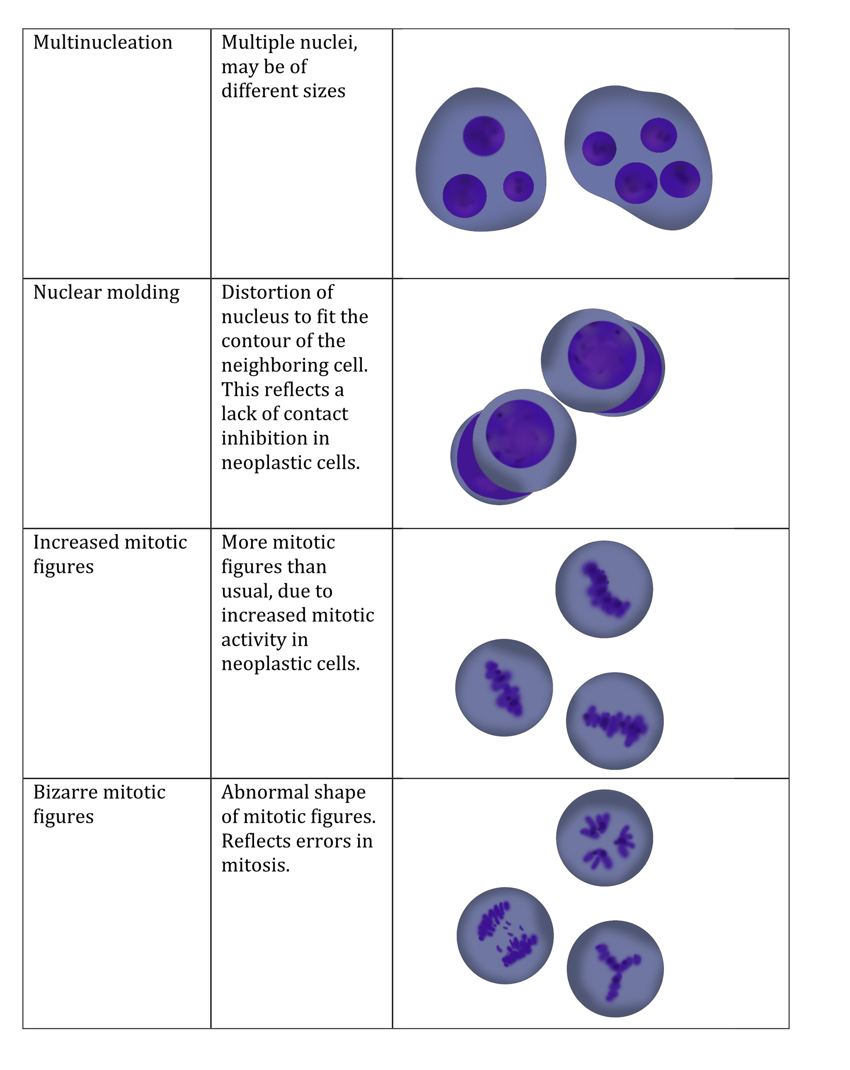
Criteria of malignancy are cytologic features that can be evaluated to help determine if a tumor is benign or malignant (Fig. 5.16). They are similar for both carcinomas and sarcomas, and include: cellular and nuclear pleomorphism (variation in cellular and nuclear morphology); anisocytosis (variation in cell size); anisokaryosis (variation in nuclear size); macrocytosis (large cells); high nuclear to cytoplasmic ratio; bizarre nuclei; multiple nuclei; macronuclei (large nuclei); prominent, large, irregularly shaped, multiple nucleoli; increased number of mitotic figures; bizarre mitotic figures; increased staining intensity. In addition, carcinomas may display nuclear molding (the ability of one cell to deform an adjacent cell; Fig. 5.17) and decreased cell-cell adhesiveness. The malignant cells represent a homogeneous or monotonous population in terms of being derived from one tissue type, despite the fact that many of the features of malignancy emphasize morphologic variability within the population. Also, some of these features are more common in certain types of tumors, and it would be very unusual to see all of the listed features within a single tumor. Carcinomas and sarcomas are occasionally so anaplastic (poorly differentiated) that it is impossible, based on cytology alone, to characterize them as epithelial or spindle cell origin. Histopathology, to examine larger numbers of cells and tissue architecture, together with additional tests such as immunohistochemistry, electron microscopy, or molecular techniques may be necessary in these situations.


Any of a variety of changes in cell morphology that are seen in malignant cells, e.g. anisokaryosis, multiple nucleoli, high nuclear to cytoplasmic ratio.
Variation in morphology.
Variation in cell size.
Variation in nuclear size.
Increased number of erythrocytes with increased volume that usually corresponds with increased MCV; may be seen in regenerative anemias and can be normal in Poodle dogs.
Larger than normal nuclei.
Distortion and displacement of the nucleus of a cell due to crowding and uncontrolled growth of neighboring cells; a feature of malignancy in epithelial neoplasms.
Poorly differentiated.


