Anemia
Anemia is an absolute reduction in the volume of erythrocytes in the peripheral blood and is identified by decreases in Hct (or PCV), Hgb, and usually RBC count, below an established reference interval (RI) for the species. See Table 1.3 which is a guideline for grading the degree of anemia in the common domestic species.

Anemias are classified in many ways, for example, according to: the responsiveness of the bone marrow (regenerative vs nonregenerative); the cause, such as blood loss, hemolysis, or bone marrow failure; the morphology of the erythrocytes, such as microcytic and hypochromic; and the etiology, such as exposure to an oxidizing agent, blood-borne parasite, traumatic hemorrhage, etc.
Erythrocyte Regeneration
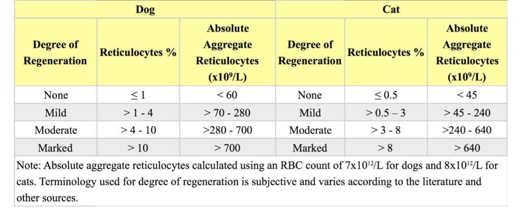
The bone marrow response to anemia, for most species, can be assessed by evaluating the peripheral blood. Blood smear examination in regenerative anemias will usually reveal macrocytosis, increased anisocytosis, and increased polychromasia. Macrocytosis relates to the release of immature erythrocytes/polychromatophilic erythrocytes and this will correlate with the MCV. Theoretically, the RDW should reflect the degree of anisocytosis, although there is not always a good correlation between the RDW and the degree of anisocytosis reported on blood smear evaluation. Increased polychromasia corresponds to an elevated reticulocyte count identified with NMB staining (see Table 1.4 for a guide to grading the response to anemia). Several species release immature erythrocytes containing many small blue dots with Romanowsky stains. This is known as basophilic stippling and is most prominent in bovine regenerative anemias, but can also be seen in other ruminants and in cats and dogs with robust regenerative responses. If basophilic stippling is not accompanied by anemia and a significant regenerative response, a disorder in hemoglobin synthesis, as seen with lead poisoning, should be considered. Metarubricytes may also be released inappropriately into the peripheral blood with lead poisoning.

Horses rarely release immature erythrocytes (polychromatophilic RBCs/reticulocytes) and nucleated RBCs (nRBCs) even when intense erythroid hyperplasia is present in the marrow in response to severe anemia. Peripheral blood findings of erythrocyte regeneration which may be, but are not necessarily, seen in the horse are increases in MCV and RDW. Bone marrow examination is required to accurately assess erythropoiesis in the anemic horse. However, the clinical information together with monitoring of the erythrogram over several days may obviate the need for bone marrow examination.
nRBCs (usually metarubricytes) may accompany a regenerative response. However, if anemia is not present, or anemia is present but nucleated RBCs are disproportionately high relative to the reticulocyte response, other conditions should be considered. These include hypoxia, necrosis, lead poisoning, acute erythrocytic neoplasia (especially in cats, see Chapter 3. Hemopoietic Neoplasia), and nonhemopoietic neoplasia metastatic to the bone marrow.
Causes of Regenerative Anemia
Regenerative anemia is usually due to blood loss or hemolysis. Following the development of anemia (e.g. due to acute hemorrhage), reticulocytes will increase in the peripheral blood by 2 to 4 days and peak by about 7 to 10 days (varying with the species). If there is no regeneration when the anemia is first detected, monitoring the peripheral blood response over several days may be necessary to determine if the bone marrow is responding appropriately.
Trauma is a frequent cause of acute external and internal blood loss. The body immediately attempts to restore the circulating volume by drawing low protein interstitial fluid into the intravascular space. Usually by the time blood is obtained for analysis, there is a decreased Hct, Hgb, RBC count, and total protein. Intravenous fluids which may be given to restore blood volume also exacerbate the drop in Hct, Hgb, RBC count, and total protein.
Increased number of erythrocytes with increased volume that usually corresponds with increased MCV; may be seen in regenerative anemias and can be normal in Poodle dogs.
Presence of small, blue, punctate inclusions in RBCs, representing aggregated ribosomes. May be seen with regenerative anemia especially in cattle, but also in dogs and cats. Occasionally seen with lead toxicity but with no or only mild accompanying anemia.
Cellular proliferation- increased number of normal cells.
Abnormal uncontrolled growth of cells that are unresponsive to normal physiologic growth controls; may be benign or malignant.

