Acute Myeloid Neoplasms
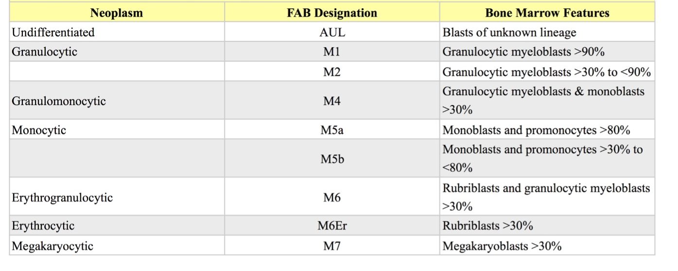
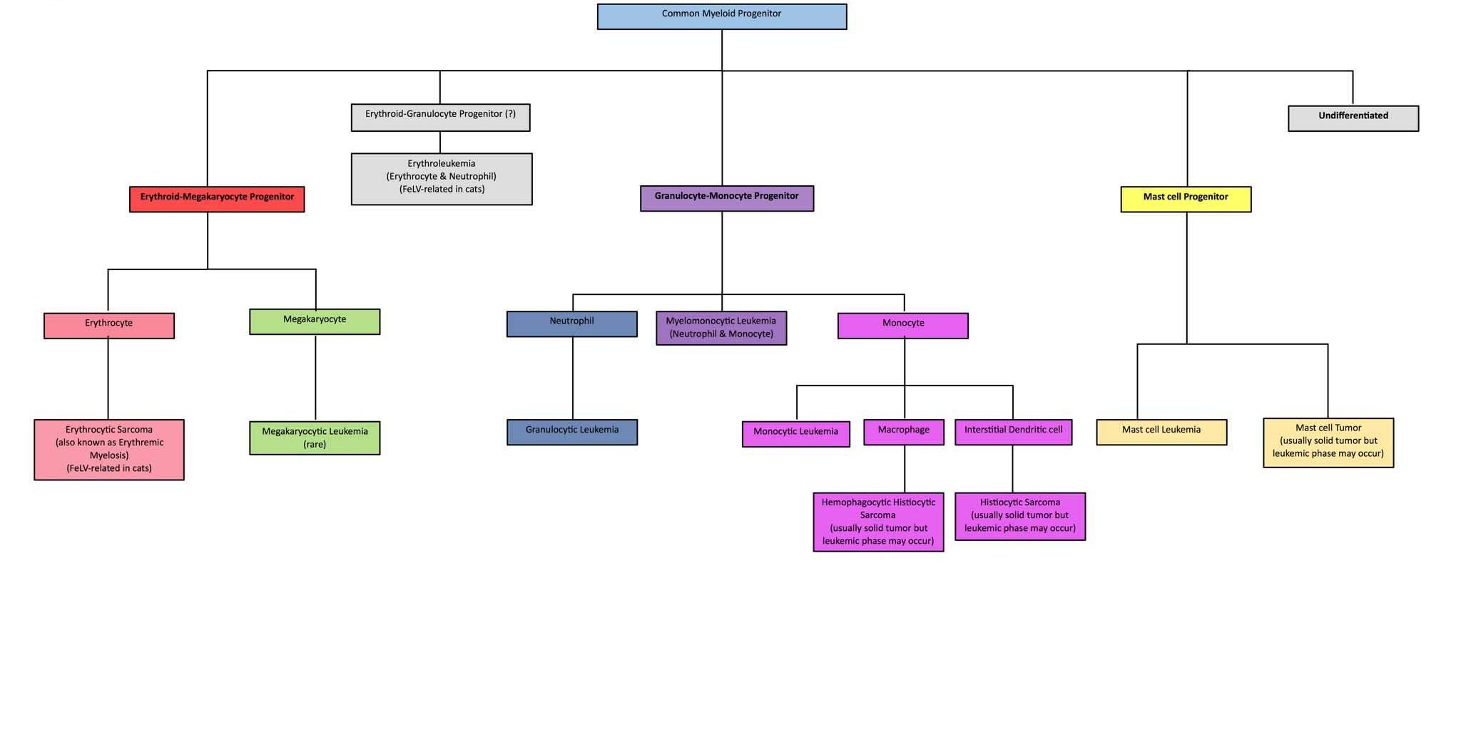
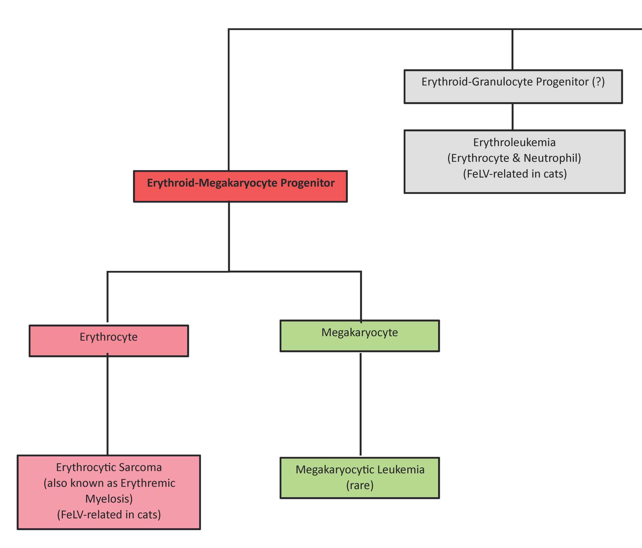
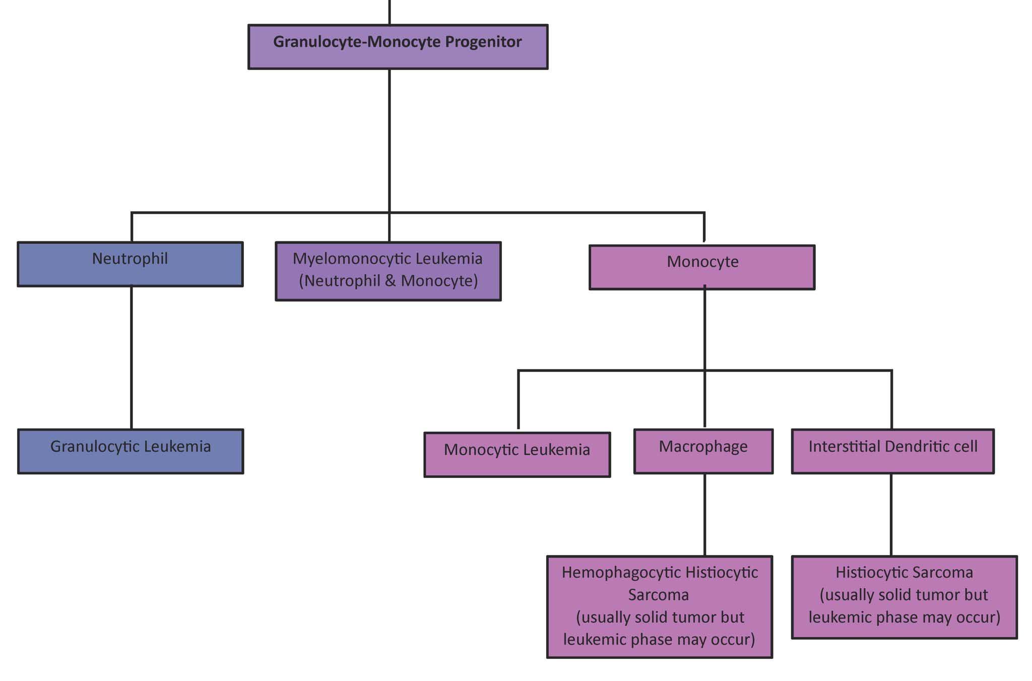
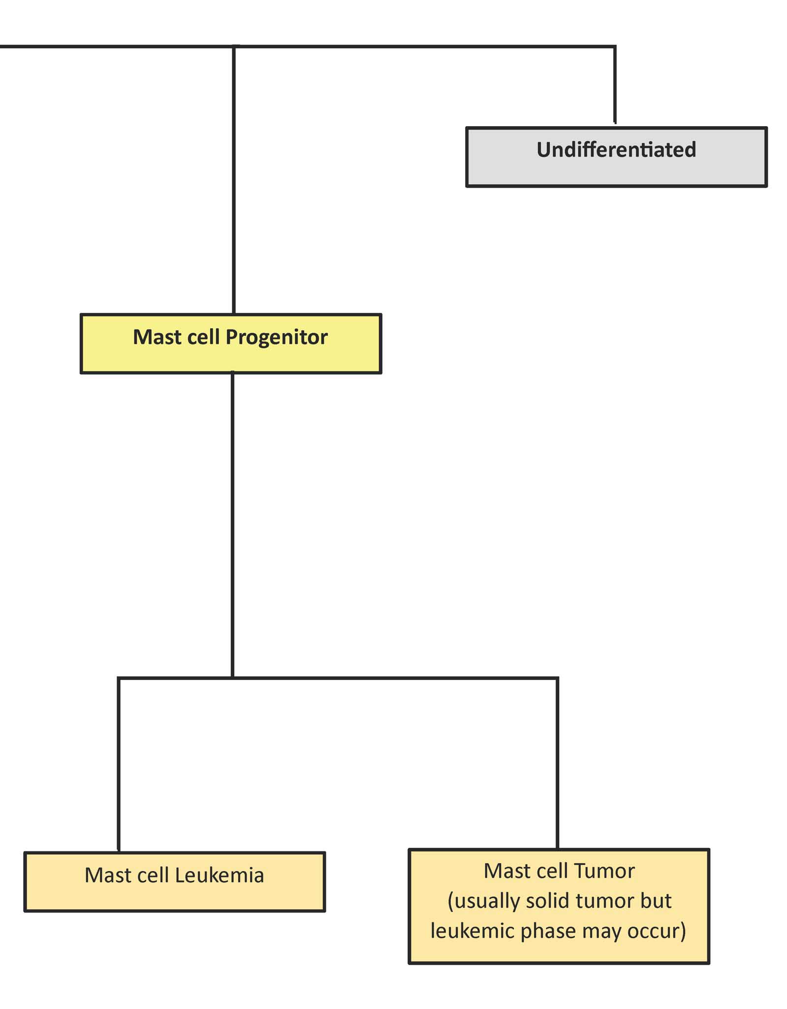
The acute myeloid neoplasms are named according to cell lineage, when known, and can be abbreviated according to the human French-American-British (FAB) classification scheme (see Table 3.2 and Fig. 3.7).
Undifferentiated
The undifferentiated category of myeloid neoplasms is reserved for situations where close to 100% of marrow cells are blasts and maturation to a specific lineage cannot be determined.
Granulocytic (M1, M2)
Granulocytic myeloblasts comprise a greater proportion of the nucleated cells in the bone marrow in M1, compared to M2. The clinical relevance of subdividing acute granulocytic neoplasia into two subtypes in veterinary medicine has not been determined. Tumors of neutrophil precursors are more common than those of eosinophils and basophils; however, specific granules are not present at the granulocytic myeloblast stage, so the line of differentiation is not necessarily known, particularly in M1. Cytochemical stains reveal variable positivity for peroxidase, Sudan black B, chloroacetate esterase, acid phosphatase, and leukocyte alkaline phosphatase.
Granulomonocytic (M4)
Cells with features of granulocytic myeloblasts and monoblasts comprise >30% of all nucleated cells in the bone marrow with granulomonocytic (also called myelomonocytic) neoplasia. In addition to cytochemical staining consistent with granulocytic lineage described for M1 and M2 above, a proportion of the neoplastic cells are also positive for nonspecific esterase. Granulomonocytic neoplasia is reported to be the most common acute myeloid neoplasm in dogs, cats, and horses.
Monocytic (M5)
The M5 category may be subdivided into M5a and M5b based on the degree of differentiation to monocytes. In M5a, monoblasts and promonocytes represent >80% of non-erythroid marrow cells. In M5b, monoblasts and promonocytes represent between 30 and 80% of non-erythroid cells, and differentiation to monocytes is noted. Cytochemical staining is usually positive for nonspecific esterase and acid phosphatase. Similar to the situation with the acute granulocytic tumors, the clinical relevance of subdividing the monocytic category is unknown in veterinary medicine.
Erythrogranulocytic (M6) and Erythrocytic (M6Er)
Differentiation of neoplastic cells along both erythroid and granulocytic lines occurs with erythrogranulocytic (M6) neoplasia, sometimes referred to as erythroleukemia. Bone marrow erythroid precursors comprise >50% of all nucleated marrow cells and granulocytic myeloblasts comprise >30% of non-erythroid marrow cells. The M6 designation can also be applied if blast cells (rubriblasts and granuloblasts) comprise >30% of all nucleated cells in the bone marrow. When the tumor involves erythroid precursors only, the M6Er designation is used and the tumor is called erythrocytic sarcoma or erythremic myelosis. Rubriblasts can be identified by experienced clinical pathologists based on morphology and staining characteristics. Also, along with rubriblasts, there are usually increased numbers of prorubricytes, rubricytes and metarubricytes, but not polychromatophilic erythrocytes. Visualizing the continuum in development from the more primitive cells to more mature cells is a very helpful tool in cell identification, in this and other tumors.
Erythrogranulocytic and erythrocytic neoplasms are seen in cats and are usually associated with FeLV infection. Abnormal maturation of marrow precursors is often present, as well as peripheral blood cytopenia(s). Delayed or asynchronous maturation of the nucleus relative to the cytoplasm (also called megaloblastic change) is commonly seen, particularly in erythroid precursors, and this leads to erythrocyte macrocytosis and increased MCV. Despite the abundance of erythroid precursors in the marrow and peripheral blood, and the increased MCV, there is little to no polychromasia or reticulocytosis (with NMB staining), so the anemia is nonregenerative and often severe.
Megakaryocytic (M7)
The M7 designation means that megakaryoblasts represent >30% of all nucleated cells in the bone marrow. Often there are increased numbers of more mature megakaryocytes in the bone marrow as well. Megakaryoblasts, and thrombocytopenia or thrombocytosis may be found on examination of the peripheral blood smear. Immunohistochemistry to identify von Willebrand factor antigen and acetylcholine esterase cytochemistry may be useful to confirm megakaryocytic lineage. Myelofibrosis is associated with many bone marrow disorders, including megakaryocytic neoplasia. Ineffective megakaryopoiesis may result in increased release of platelet-derived growth factor (PDGF) which stimulates fibroblast growth and proliferation.
Stage in granulopoiesis following the committed stem cell stage; earliest form recognizable as a granulocyte precursor.
Granulocyte with fine, inconspicuous cytoplasmic granules and a segmented nucleus; important in phagocytosis and killing of bacteria.
See Secondary granule.
White blood cell (WBC); includes neutrophils, eosinophils, basophils, monocytes, lymphocytes, mast cells.
Stage in erythropoiesis following the committed stem cell or CFU-E stage; earliest form recognizable as an erythrocyte precursor.
Stage in erythropoiesis following rubriblast.
Stage in erythropoiesis following prorubricyte; hemoglobinization is most active during this stage.
Stage in erythropoiesis following rubricyte and the final stage of erythrocyte development to possess a nucleus in mammalian species.
Polychromatophil; an anucleate (in mammalian species), immature erythrocyte containing cytoplasmic RNA and ribosomes which give the cell a bluish-pink appearance with Romanowsky stains (e.g. Wright-Giemsa).
Increased number of erythrocytes with increased volume that usually corresponds with increased MCV; may be seen in regenerative anemias and can be normal in Poodle dogs.
Average erythrocyte size in femtoliters (measured or calculated PCV or Hct÷RBC).
Increased numbers of anucleate, immature erythrocytes that stain bluish-pink with Romanowsky stains due to the presence of cytoplasmic RNA.
Decrease in hematocrit (PCV) recognized on the complete blood count (CBC); usually hemoglobin concentration and RBC numbers are also decreased.
Stage in megakaryopoiesis following promegakaryoblast.
Decrease in the number of circulating platelets below the reference interval.
Protein important in binding platelets to subendothelial collagen.
Part of hemopoiesis dealing with the production of megakaryocytes.

