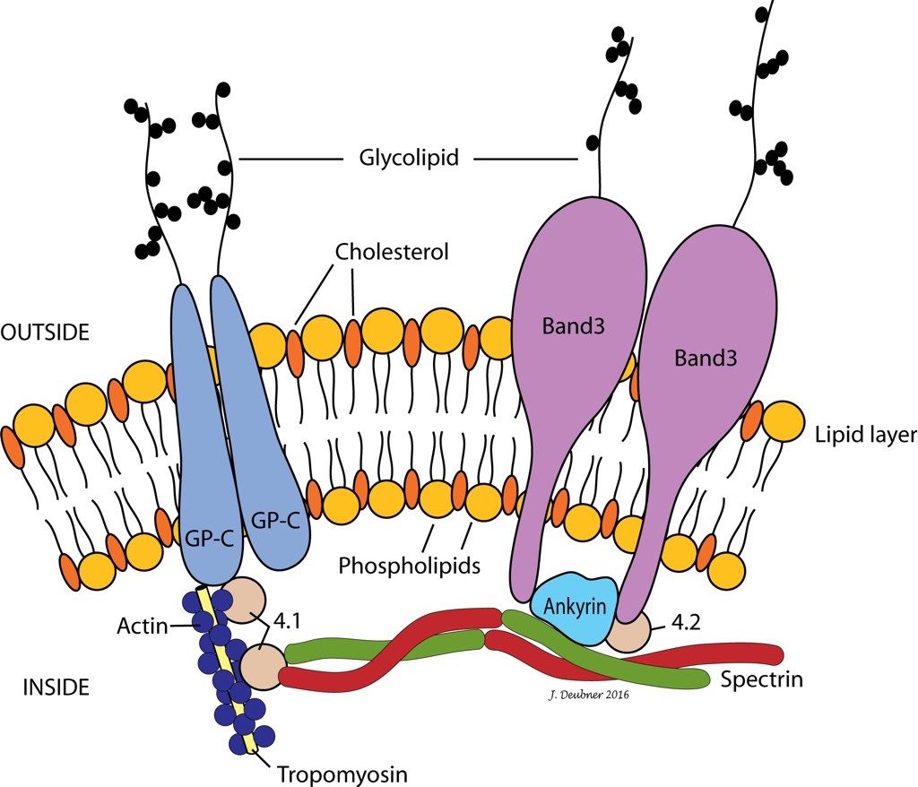
Erythrocyte Membrane and Cytoskeleton
Erythrocyte Membrane
The red cell membrane is a typical lipid bilayer, comprising mainly phospholipids and cholesterol (Fig. 1.30). Membrane proteins and glycoproteins are inserted into the lipid bilayer, some in one leaflet only and others spanning the entire membrane. Carbohydrate residues expressed by glycoproteins on the outer RBC surface contribute negative charge and antigenic determinants. The integral membrane proteins include receptors and enzymes, which usually only partially penetrate into the bilayer, and transmembrane proteins, which include the transport carriers for ions and water soluble substrates, such as glucose. The transmembrane proteins maintain cation and anion homeostasis within red cells. Abnormalities in ion pumps can result in a shortened red cell lifespan.

Erythrocyte Cytoskeleton
The RBC cytoskeleton forms the scaffolding for the lipid bilayer and plays an important role in membrane stability and deformability. Deformability is paramount for survival of RBCs, particularly as they maneuver physically challenging capillary beds, such as those within the spleen. Cells that are not deformable are subject to aggressive removal by the mononuclear phagocyte system (MPS). Interactions between skeletal proteins such as spectrin, ankyrin, and actin and the lipid bilayer, maintain membrane shape, flexibility, and durability. Interactions between skeletal proteins and transmembrane proteins stabilize the organization of membrane phospholipids and influence cell surface activities (e.g. anion and glucose transport). RBC skeletal proteins link to the lipid bilayer via two main interactions: spectrin-ankyrin-band 3 and spectrin-actin-band 4.1. The bands are named according to their migration patterns on electrophoresis, but many are now assigned names which reflect their function. For example, band 3 is now known as AE1 for its role as an anion exchange protein or channel. Defects in skeletal proteins can result in abnormal erythrocyte morphology, sometimes associated with increased membrane fragility and decreased RBC survival. In other cases, although the abnormal erythrocyte morphology may be readily seen on peripheral blood smear examination, clinical effects may be absent.
General term for fat, including triglycerides, phospholipids, cholesterol.
Type of lipid used to form cell membranes.
Lipid used to form cell membranes, steroid hormones, bile acids, and vitamin D.
Group of phagocytic cells mainly derived from monocytes and differentiating to macrophages in various tissues.
Erythrocyte cytoskeletal protein.
Erythrocyte cytoskeletal protein.
Erythrocyte cytoskeletal protein.
Erythrocyte transmembrane protein; now called AE1, anion exchange channel
Erythrocyte cytoskeletal protein

