Evaluation of Fluid Samples
Fluid samples obtained for cytologic examination include: pleural, peritoneal, and pericardial fluid; synovial fluid; cerebrospinal fluid; ocular fluids (aqueous humor and vitreous humor); and bile. Samples from fluid-filled masses can also be submitted for preparation of concentrated smears. Although exceptions exist, complete evaluation comprises: gross appearance (i.e. color and clarity), quantity of fluid submitted, total protein concentration, nucleated cell count, red blood cell count, and microscopic examination. Microscopic examination of smears of fluid samples is similar to that for samples from solid tissues (described above).
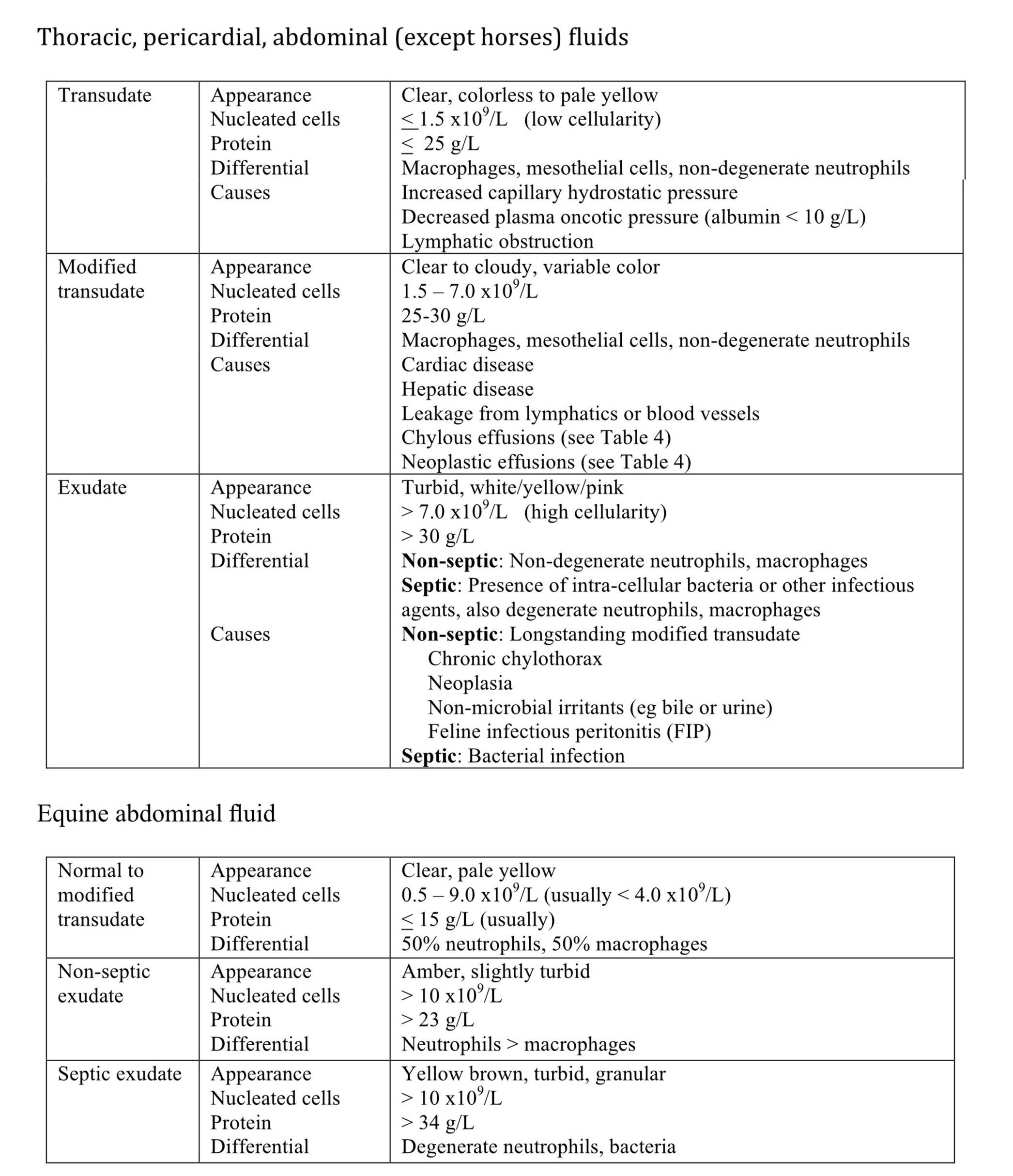
Thoracocentesis or abdominocentesis of normal animals produces little or no fluid, with the exception of the horse from which about 5 mL of yellow abdominal fluid can usually be obtained. Pleural and peritoneal fluids are categorized according to the protein concentration and nucleated cell count. Guidelines for interpretation vary in the veterinary literature and there is a degree of overlap across the three categories (Table 5.2). However, determining the type of fluid can help to formulate the differential diagnoses in many situations.

Process of obtaining thoracic fluid for cytologic evaluation.
Process of obtaining abdominal fluid for cytologic evaluation.

